An AI model accurately predicts how cells end up in position inside tissues
Called TopoVelo, the model could lead to better understanding of cellular interactions
5:00 AM
Authors |
How do cells come together to make tissues?
To answer this question, the laboratory of Joshua Welch, Ph.D., has developed an A.I. powered computational tool that incorporates space and time into models of cell fate transition.
This modeling, described in a recent issue of Nature Biotechnology, constitutes a key step toward characterizing how interactions and environment among neighboring cells and cell migration contribute to tissue development.
“Previous studies have modeled cell differentiation like a physical process in which a cell is a point mass moving through space with some velocity. But cells exert forces on each other much like bodies in space interact through gravity and momentum transfer,” explained Welch, an associate professor of computational medicine and bioinformatics at the Medical School, and principal investigator for the study.
“Inspired by this analogy, we used artificial intelligence to model how the gene expression levels of cells from an entire tissue influence each other while the cells differentiate.”
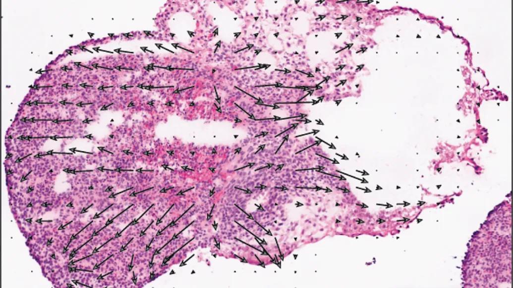
Called Topological Velocity Inference (TopoVelo), the Welch model jointly infers how cells change in location over time from data detailing where genes are expressed in tissue.
“Previous research has measured the direction and speed that cells are moving, but cells are not moving alone: they are all bumping into each other and pulling on each other, much like gravity pulls planets together,” he explained.
The Welch lab used TopoVelo, a type of graph neural network, to estimate cell velocity from the developing mouse cerebral cortex and found that it learns the location and maturity of cells based on the expression of ligand-receptor genes–genes that facilitate the connection of one molecule to another–revealing spatial signatures of mouse neural tube closure, a critical point of early development.
Extending the model to humans, the Welch lab generated Slide-seq data, which reveals where genes are expressed in a tissue, from an in vitro model of human development and used TopoVelo to study the spatial patterns of early differentiation.
“There are many diseases, such as neuronal migration disorders, that evolve from incorrect cell migration and models like this provide a way to determine what’s causing migration defects and how we could potentially correct them,” Welch said.
The A.I. model could also provide a better understanding of the cell-cell interactions underlying inflammatory responses.
Additionally, said Welch, TopoVelo could provide the insight to engineer better organoids by more correctly modeling how cells under investigation differentiate.
Additional authors: Yichen Gu, Jialin Liu, Kun H. Lee, Chen Li, Lu Lu, Jaimee Moline, Renxiang Guan
Funding/disclosures: National Institutes of Health grants R01 HG010883 and UM1 MH130966
Paper cited: “Topological velocity inference from spatial transcriptomic data,” Nature Biotechnology . DOI: 10.1038/s41587-025-02688-8

Health Lab
Explore a variety of health care news & stories by visiting the Health Lab home page for more articles.
Media Contact

Public Relations
Department of Communication at Michigan Medicine
In This Story

Joshua Welch, PhD
Associate Professor
Stay Informed
Want top health & research news weekly? Sign up for Health Lab’s newsletters today!
Featured News & Stories
Studying neurons using neurons

The algorithm will see you now? Patients say not without a doctor nearby
How AI is helping emergency physicians learn from their patients
Research may help better predict outcomes in kids with congenital cytomegalovirus

Michigan’s aging brains need more protection, poll shows