Big ideas. Big leaps forward.
9 Michigan Answers that could change the world … and your life
Author |
With additional reporting by Tara Roberts and Kate Murphy and illustrations by Ellen Weinstein
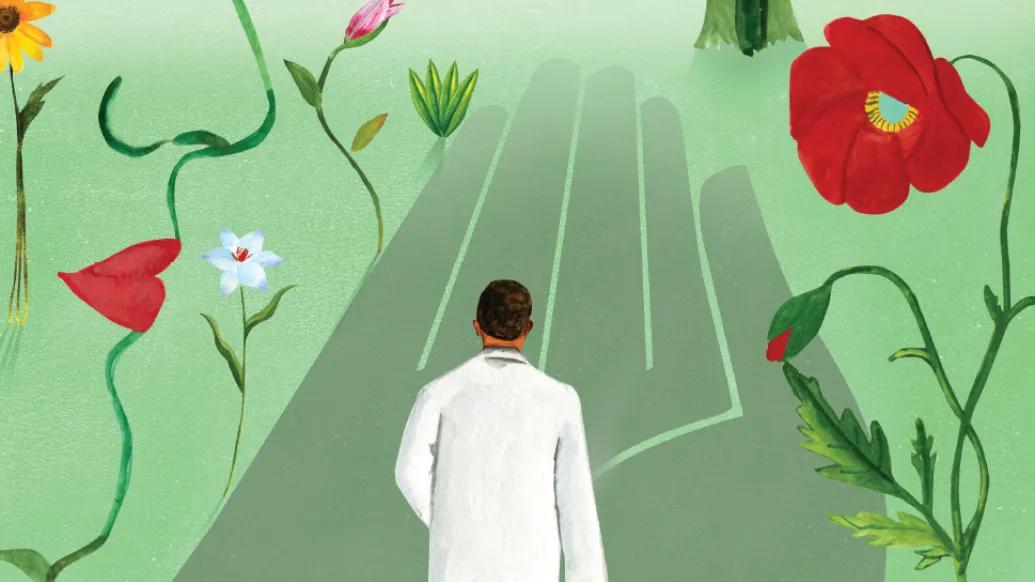
Being a good researcher requires a special combination of skills. You’ve got to be pretty good at grunt work: applying for grants, collecting and analyzing data, and doing administrative tasks, like organizing labs and assistants. But you also need an excellent imagination. You must be able to envision brighter days ahead and formulate a question that, if answered, could lead humanity to that future.
Many researchers at Michigan Medicine are the kind of visionaries who could help us across the bridge between science fiction and reality. In this story, we feature just a handful of those researchers who are undertaking projects inspired by big dreams. They’ve imagined a future where we catch cancer before it metastasizes, where we diagnose bipolar disorder with a simple blood test, where there’s no functional difference between prosthetic and biological limbs, and several other possibilities for making our health, and our lives, better.
Jump to a section of this article:
A blood test to diagnose bipolar disorder?
Buying more time for an injured soldier?
Body odor to the rescue?
It has been well known since Hippocrates that many diseases have distinct odors associated with them, such as the fruity smell that accompanies diabetic ketoacidosis,” says Xudong (Sherman) Fan, Ph.D., the Richard A. Auhll Professor of Engineering at U-M, professor of biomedical engineering, and associate director of the U-M Max Harry Weil Institute for Critical Care Research and Innovation.
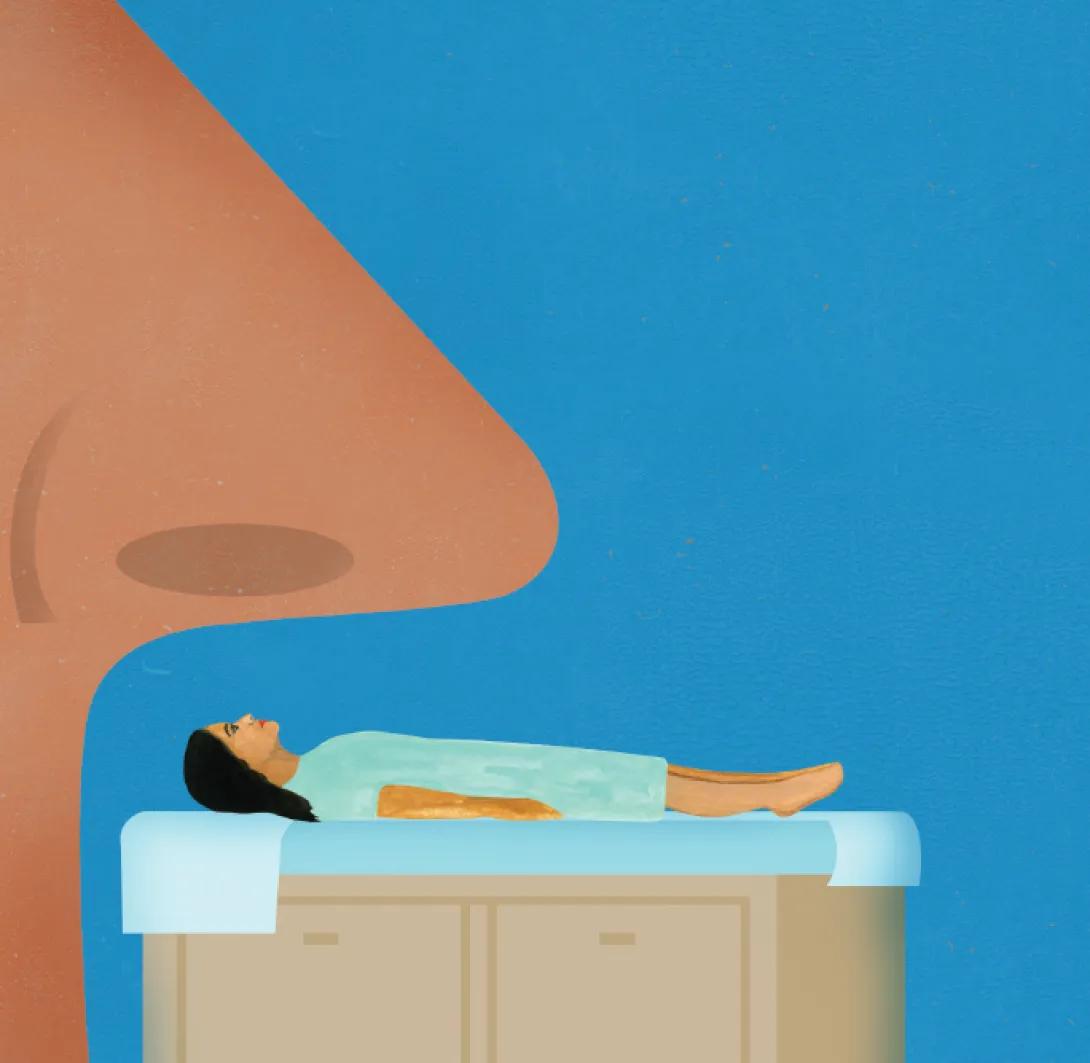
“These odors are the result of volatile organic and inorganic chemical compounds emanating from the skin, which are reflective of the human body’s metabolic processes as well as the bacteria and viruses living within it. Analyzing these compounds could provide us with unique diagnostic clues, guide laboratory evaluation, and facilitate and expedite treatment.”
Fan is leading a group of researchers who received a $5.7 million grant from the NIH Screening for Conditions by Electronic Nose Technology (SCENT) program to develop a portable sensor that uses body odor to detect more than 20 diseases in both adults and children.
Currently, few technologies exist for wearable body odor analysis. Benchtop gas chromatography devices are bulky and impractical. E-nose sensors, while simpler and faster, are susceptible to environmental changes and can suffer from crosstalk among their various sensing elements. By combining existing technologies with advanced machine learning and artificial intelligence algorithms, the team aims to surpass current body odor analysis methods and greatly enhance their device’s pattern recognition and disease detection capabilities.
The team hopes the device will be able to detect everything from critical illnesses like sepsis, stroke, congestive heart failure, GI bleeding, and diabetic ketoacidosis to skin diseases (such as psoriasis) and pulmonary diseases (such as asthma and COPD). Recently the Weil team demonstrated the ability of a breath version of the approach to diagnose acute respiratory distress syndrome (ARDS), the most life-threatening form of lung injury. It can also diagnose COVID-19.
Artificial ovaries?
Treatment for leukemia has improved dramatically in the last couple of decades — in children, this cancer is now considered almost curable. But the side effects of treatment can still be harsh. One such side effect for pre-adolescent female patients is premature ovarian insufficiency. This not only causes sterility but also symptoms related to the disruption of ovarian endocrine function, such as loss of bone density, muscle wasting, impaired cognitive development, and accelerated cardiovascular disease.
Ariella Shikanov, Ph.D., associate professor of biomedical engineering and of obstetrics and gynecology, aims to fix that.
Just as with other organ transplants, the transplanting of ovarian tissue comes with the risk of rejection. The researchers in Shikanov’s lab have figured out a way around that risk. By using immunoisolating capsules, made with hydrogels, they may be able to give patients functioning “ovaries” without the patient’s own immune system attacking the ovarian tissue that comes from a donor. The researchers have already achieved this in a mouse model and are on track to start clinical trials in humans by the end of the year.
Although Shikanov’s project started out to help a relatively small group of patients, the research has the potential to improve the lives of many more. Hormone replacement therapy (HRT) for people who have gone through menopause carries with it an increased risk for blood clots, stroke, and breast cancer, among other side effects.
“HRT doesn’t talk to fat, to muscle; it just delivers estrogen and floods the whole system,” Shikanov says. Though there is not yet evidence to show that artificial ovaries would be better, Shikanov believes it’s possible that having hormones that come organically from ovarian tissue could reduce or eliminate those side effects.
A blood test to diagnose bipolar disorder?
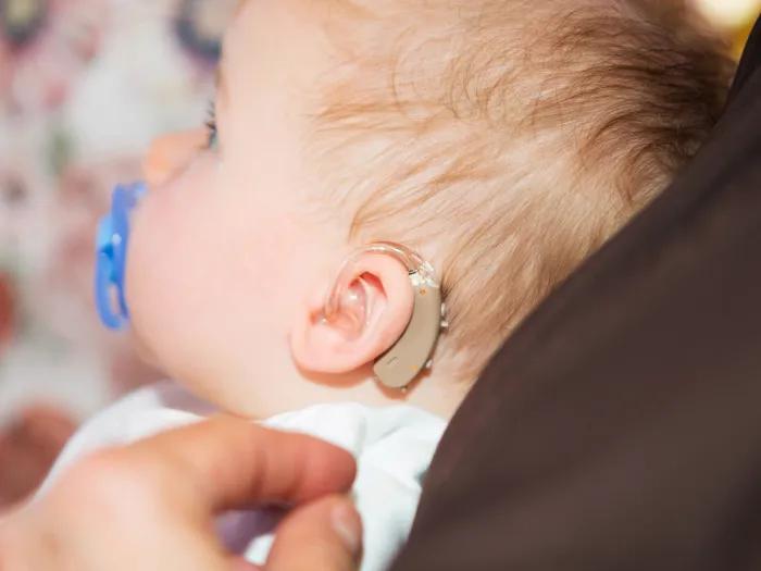
Are the brain cells of a person with bipolar disorder different from other people’s cells?
They appear to be, according to research in the lab of Sue O’Shea, Ph.D., the Crosby-Kahn Collegiate Professor of Cell & Developmental Biology and professor of psychiatry. O’Shea is a member of the Heinz C. Prechter Bipolar Research Program.
The researchers took skin cells from people with bipolar disorder and from individuals not diagnosed with psychiatric illness, turned back their biological clock until the skin cells behaved like pluripotent stem cells, and then studied their behavior. There was a striking difference between the cells of the two groups, particularly in the way they passed signals from cell to cell.
When they compared the expression of proteins in the supporting cells (astrocytes), they found eight proteins that were significantly overexpressed in individuals with bipolar disorder compared with controls, O’Shea says.
There are several implications of their discovery. The most exciting is the possibility of finding a druggable target among those proteins to treat bipolar disorder. For example, O’Shea says some of those overexpressed proteins have been associated with neurodegenerative diseases. “If there is a component of a degenerative process in bipolar disorder, it might be possible to repurpose drugs already on the market for neurodegeneration to treat bipolar disorder.”
Another possible outcome of O’Shea’s research would be a diagnostic tool, such as a blood test, that would allow for earlier intervention.
O’Shea hopes the research may also help destigmatize mental illness. She has seen individuals with bipolar disorder look through the microscope and say, “See, my cells are different.”
Regrow your own knee tissue?

Knee injuries happen. In fact, millions each year result in visits to a doctor. Nearly 350,000 of those injuries require surgery to reconstruct the anterior cruciate ligament (ACL).
ACL reconstructions typically involve bone/ligament autografts (with tissue taken from other parts of the patient’s body) or allografts (with donated tissue), both of which have major downsides. The patient’s knees are often not restored to full function. And there’s a risk of tissue rejection with allograft repairs.
A group of Michigan Medicine researchers who co-founded STEL Technologies, LLC — Lisa Larkin, Ph.D., professor of molecular and integrative physiology and of biomedical engineering; Ellen Arruda, Ph.D., the Tim Manganello/BorgWarner Chair of Mechanical Engineering, the Maria Comninou Collegiate Professor of Mechanical Engineering, and professor of biomedical engineering; and Edward Wojtys, M.D., the William S. Smith Collegiate Professor of Orthopaedic Surgery — have created a solution in the form of stem-cell generated ACL grafts.
The cell-generated extracellular matrix (CGEMTM) graft they’ve created is a devitalized combination tissue consisting of two mineralized (bone) areas and a non-mineralized (ligament) area with extracellular matrices made of mesenchymal stem cells (a type of stem cell found in bone marrow). In a sheep model of ACL repair, STEL’s graft was able to restore near normal form and function to an ACL-injured knee and eliminate the donor site pain and disability associated with autografts, additionally overcoming many of the shortcomings related to graft failure and other risks of allografts.
The CGEMTM graft is fabricated at a slightly larger length than is needed, and upon implantation, it re-establishes tension by contracting, providing a patient-specific fit. It doesn’t require fixation screws. And it allows the patient’s body to regrow native ACL tissue.
Manufactured by STEL Technologies, LLC, the graft will be ready for first-in-human clinical trials within 12 months.
Inventing a new vital sign?
Pulse rate. Blood pressure. Temperature. Respiratory rate. When you’re ill or injured, a health care provider will measure these four traditional vital signs to determine how serious your condition is. There’s just one problem: The traditional vital signs may not change until it’s too late to get help, especially if you have an illness like pneumonia, a blood infection like sepsis, or trauma. Traditional vital signs aren’t great at predicting if you will become hemodynamically unstable — that is, develop low blood pressure, which can lead to inadequate blood flow to vital organs.
A team at the Max Harry Weil Institute for Critical Care Research and Innovation has developed a new analytic to examine a simple single electrocardiogram (ECG) signal. They discovered that there are changes in the ECG that are imperceptible to the human eye that predict, with high accuracy, if patients will become hemodynamically unstable — sometimes hours before the episode occurs. This provides enough time for health care providers to intervene and prevent complications, including death.
The team created a product called the Analytic for Hemodynamic Instability (AHI) that monitors the autonomic nervous system (ANS), which helps control the cardiovascular and respiratory systems. AHI enables detection of ANS dysfunction, which occurs before overt changes in blood pressure happen, essentially providing a new early vital sign to assess.
Licensed to U-M start-up Fifth Eye, Inc., AHI recently got FDA approval for predictive monitoring and is being used in hospitals around the country. The technology is designed to be used with ECG monitoring, including wearable ECG patches, which may have a tremendous impact for monitoring patients no matter where they are — from soldiers on the battlefield to patients at home.
Predicting preterm birth?
The State of Michigan got a D+ from the March of Dimes in the organization’s most recent national maternal and infant health report card. The state’s preterm birth rate climbed from 10.2% to 10.6% from 2020 to 2021.
In the U.S., more than 10% of babies are born preterm. They face not only immediate risk of death but also lifelong risks of adverse health outcomes.
“We do not understand the timing of birth,” says Molly Stout, M.D., the Morton R. Lazar Professor of Obstetrics and Gynecology Innovation. “I can see a patient in clinic today, and she’ll go into labor tomorrow. And I wouldn’t be able to tell you today that that was going to happen.”
And that information is knowable. That progress is achievable.”
A device that can more accurately predict preterm birth can help people receive critical interventions to improve neonatal survival. It could also help avoid unnecessary hospitalizations and interventions.
Stout has assembled a team of investigators — including physicians, sonographers, and engineers from U-M, Brown University, and Washington University in St. Louis — to find a way to predict preterm birth. They performed more than 1,100 cervical imaging visits using a novel ultrasound-based device that measures how soft or firm the cervix is. They were able to detect the cervical softening that occurs before the cervix dilates for birth and are now testing whether the device can be used to predict who is at the highest risk to deliver early.
The group will begin developing novel cervical imaging this year which will measure both the length of the cervix and how soft or firm it is. Once refined, the technology could allow providers to determine if certain preterm birth prevention strategies are working during pregnancy, rather than waiting until women deliver preterm or at term.
Buying more time for an injured soldier?
When a soldier is shot in the abdomen, medics can’t use a tourniquet or apply direct pressure like they would to a wounded leg. If that soldier is in an isolated war zone, it could take up to 72 hours to evacuate.
Sometimes, a catheter is placed in the leg and snaked up to the aorta where a balloon is inflated to stop the bleeding. However, this technique requires special training and is not widely available on the battlefield.
A team at the U-M Max Harry Weil Institute for Critical Care Research and Innovation, including clinicians with U.S. military combat deployment experience, have designed a device that solves the problem less invasively. The GROA (gastroesophageal resuscitative occlusion of the aorta) takes advantage of the stomach’s position next to the abdominal aorta. The GROA device is a tube with a balloon on the end that goes into the mouth, down the esophagus, and into the stomach. By inflating a balloon and swelling the stomach, enough pressure can be placed on the aorta to “pinch” it closed, stopping the hemorrhage while maintaining blood flow to the heart and brain.
The technology is designed to be easily performed by a combat medic or civilian paramedic and could help not only soldiers but also anyone with internal bleeding from motor vehicle accidents or gunshot wounds who cannot get to an operating room quickly.
The device has been hugely successful in improving survival in preclinical models of lethal hemorrhage. It is currently undergoing the next stage of development to prepare it for clinical trials. It was recently licensed by a U-M spin-off company, Precision Trauma LLC (a veteran-owned and operated small business) for commercialization.
Mind-reading prosthetics?
A defining moment in the history of bionic limbs happened at a café near Paul Cederna’s lab in May 2022. For the first time, a patient outside the lab tested an advanced prosthetic hand controlled by her own nerve signals.
“She was using her prosthetic to get her soft drink. She was using her prosthetic to carry the bag with her food in it,” says Cederna, M.D., the Robert Oneal Collegiate Professor of Plastic Surgery and professor of biomedical engineering.
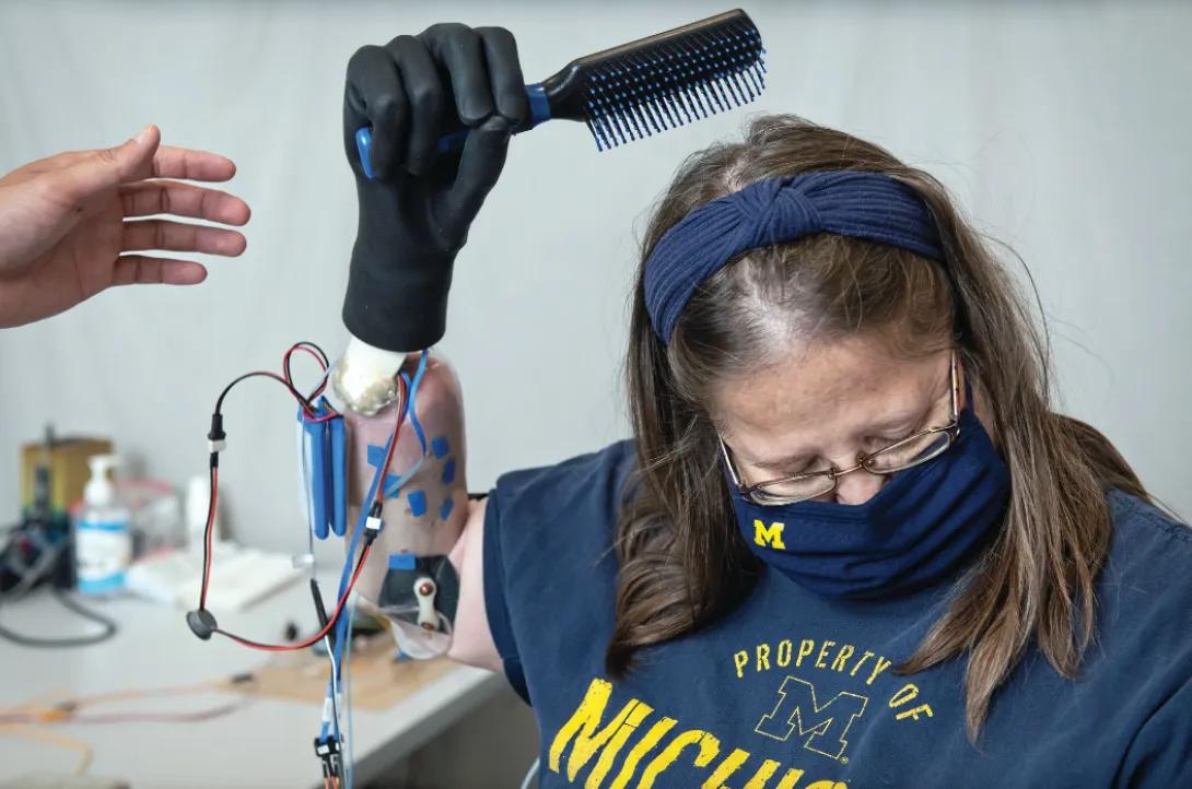
The patient, Karen Sussex, had her right hand amputated in 2017 after sepsis. In 2018, she became the first patient to participate in the clinical trial led by Cederna and Cynthia Chestek, Ph.D., associate professor of biomedical engineering.
High-fidelity finger control is the most remarkable result of the U-M technique — things like pinching clothespins and making coffee. In the past, researchers used electrodes to attempt to pick up nerve signals and translate them to prosthetic control.
“The problem with that is the signals in nerves are really tiny, and the electrical noise in the body is really big,” Cederna says.
Cederna’s alternative technique involves wrapping a tiny piece of muscle around the end of a nerve in the arm or leg, which can cause this small muscle to contract in the same way the muscle used to contract prior to losing the hand.
“Instead of recording nerve signals, which are tiny, we now can record signals from that piece of muscle that are really amplified,” he explains. “Now we know exactly what the brain is saying … That was the game changer.”
The technique — called regenerative peripheral nerve interface, or RPNI — is already helping people with limb loss around the world. Even without connecting to a prosthetic limb, the RPNI reduces phantom pain and neuroma pain. Cederna alone has performed the surgery for hundreds of patients.
Implantable cancer catcher?
Nearly 30% of women diagnosed with early-stage breast cancer develop metastatic disease. But what if you could catch those metastatic cells before they migrate to other parts of the body?
Researchers at Michigan Medicine have created an implantable scaffold that does just that. The device, which resembles a tiny sponge (5 mm in diameter and 2 mm thick), is inserted subcutaneously, and captures metastatic tumor cells before they can colonize their organ targets.
Using a needle biopsy, clinicians can look for tumor cells in the scaffold directly, or alternatively can look for immune cells that are responsible for recruiting tumor cells to solid organs. If metastatic cells or premetastatic tissue is detected, a patient could start treatment prior to the progression of disease.
Lonnie Shea, Ph.D., the Steven A. Goldstein Collegiate Professor of Biomedical Engineering, and Jacqueline S. Jeruss, M.D., Ph.D., professor of surgery and associate vice president for research, have led the development of the implantable scaffold, and are working with the Coulter Translational Research Program to obtain FDA approval for a first-in-human clinical trial, beginning in 2023. Together, they are optimistic that, with more research, the technology could be used to screen for several types of metastatic disease, including lung, pancreatic, prostate, and ovarian cancers as well.
Note: Innovative research projects are often supported by start-up companies and other groups. Though some are listed in this article, below is a full list of disclosures.
Body odor to the rescue?
The University of Michigan and Xudong Fan, Ph.D., have financial interests in Arborsense, Inc.; Blu Biotech, Inc.; and ChromX Health Co., Ltd. Dr. Fan has filed for patents on the technology and has received a patent related to COVID-19 detection.
Artificial ovaries?
Ariella Shikanov, Ph.D., holds a patent for artificial ovaries technology, which was issued in February 2021. She is also the founder and CEO of ArtOva Therapeutics, Inc., a startup company launched to translate this technology to humans.
Regrow your own knee tissue?
Lisa Larkin, Ph.D., Ellen Arruda, Ph.D., and Edward Wojtys, M.D., co-founded STEL Technologies, LLC, to commercialize their cell-generated extracellular matrix (CGEMTM).
A test to diagnose bipolar disorder?
No conflicts of interest to report.
Inventing a new vital sign?
The Analytic for Hemodynamic Instability technology is licensed to U-M start-up Fifth Eye, Inc.
Implantable cancer catcher?
Lonnie Shea, Ph.D., and Jacqueline S. Jeruss, M.D., Ph.D., have filed for patents on their scaffold application, and they are in various stages of the patent process.
Buying more time for an injured soldier?
The GROA (gastroesophageal resuscitative occlusion of the aorta) device has been licensed by a University of Michigan spin-off company, Precision Trauma, LLC, for commercialization.
Mind-reading prosthetics?
Paul Cederna and his team have obtained a series of patents and this year started a company, Blue Arbor Technologies, Inc., to produce all products and software related to this work and control neuroprosthetic devices.
Predicting preterm birth?
A patent has been filed for this work.
Related
Turning "a-ha" moments into big breakthroughs
Featured News & Stories

Researchers create new path to target hard-to-drug prostate cancer protein

How new care models, within a new building launch, are helping optimize patient care
Path forward for glioblastoma treatment

Studying neurons using neurons
How AI is helping emergency physicians learn from their patients