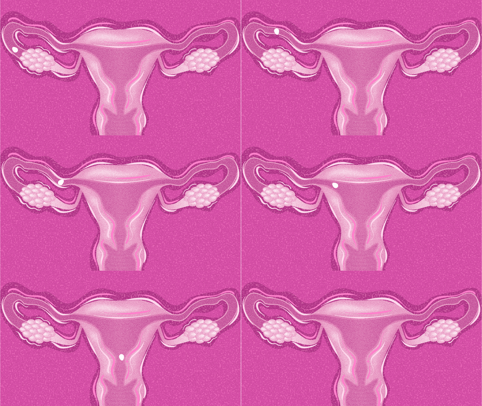
Studying human development in a dish
A new technique could provide powerful insights into early cell differentiation.
5:00 AM
Author |
Sperm meets egg, setting off a cascade of events leading to the creation of an embryo. But how do developing cells—which all contain the same genetic information—decide what to be when they grow up? How does a cell differentiate into muscle, skin, organs, or even new eggs and sperm?
New research may hold clues and provides surprising insights into the development of primordial germ cells—the cells that eventually mature to enable reproduction.
"The question of how primordial germ cells arise in humans is hard to study because there is no good experimental system to use," said Idse Heemskerk, Ph.D., assistant professor in the Department of Cell and Developmental Biology.
Much of the research to date has centered on animal models, which are imperfect models for human development. Alternative studies using cells in a dish have also had mixed results. Such in vitro studies, explained Heemskerk, begin with human derived pluripotent stem cells—stem cells modified to become embryonic-like with the inherent potential to generate any cell in the body. These cells are then grown and observed over a period of days.
However, "stem cell differentiation processes are poorly controlled, and primordial germ cells might be one of the worse," explained Heemskerk. "The yield varies a lot from cell-line to cell-line and from experiment to experiment."
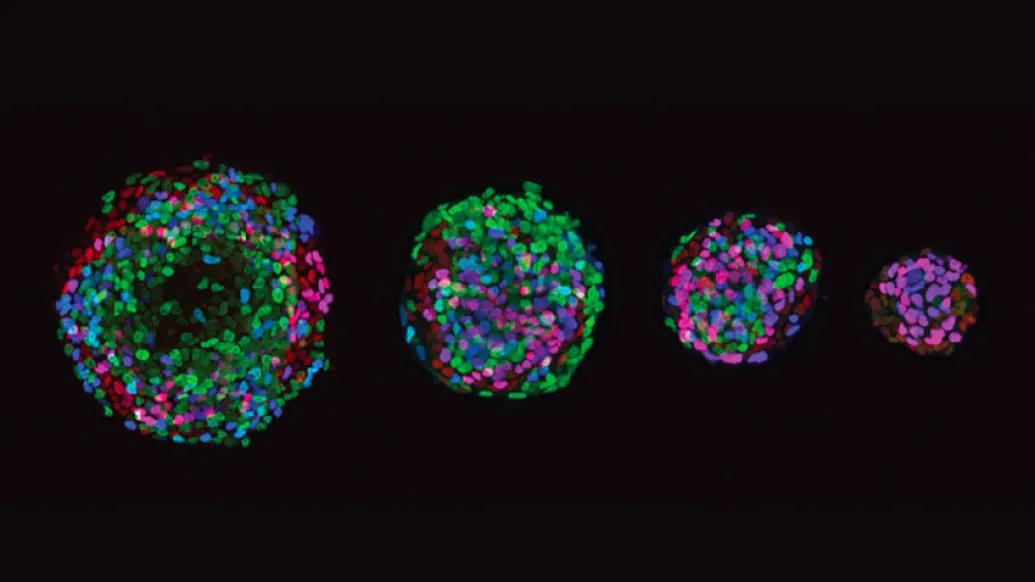
In a paper published in the journal eLife, Heemskerk, research fellow Kyoung Jo, Ph.D., and their team describe a powerful new model system that uses a technique called micropatterning to bring some control to the chaos. With the technique, the lab dish is modified so that cells can only stick to certain areas. This results in a more organized pattern of stem cell differentiation.
"If you control the number of stem cells and the way they are arranged, then the things they do become much more reproducible and when they do the same thing every time you can actually dissect what is happening," he said.
In the micropatterned dish, pluripotent stem cells reliably form concentric circles of different cell types that in the human embryo arise during a process called gastrulation, explains Heemskerk. The cells are divided up into three lineages of the body: an outer layer that becomes skin and nerves, an inner layer that becomes the gut and organs and a layer in between that makes muscle bone and blood. Visible also in these new in vitro models is a fourth cell type—the primordial germ cells that become reproductive cells.
Heemskerk explains that these cells were previously assumed to be gut cells because in mice, they expressed a marker that can only be found in the mouse gut.
Furthermore, the model enabled their team to determine why cells were aligning the way they were in a methodical way.
"Cells send signals to each other to coordinate who does what…and we have drugs to block those signals and see what happens."
One such signal, called BMP, was thought to set off a cascade for the development of the embryo by turning on another signal called WNT, which in turn, activated a different signal called Nodal.
By blocking each step of that cascade, the team was able to identify that Nodal, not WNT as commonly believed, was most important for the development of primordial germ cells.
They also found that primordial germ cells always developed at a fixed distance from the edge of the dish. By making the micropatterned area smaller, they could more than double the yield of germ cells—a critical development for researchers working on the basic science of infertility.
Said Heemskerk, "We are trying to understand the basic principles of development and the signals that define the different cell types. We have a system now where we can better study how the human germ line arises."
Paper cited: "Efficient differentiation of human primordial germ cells through geometric control reveals a key role for Nodal signaling," eLife. DOI: 10.7554/eLife.7281
Health Lab
Explore thousands of health news & research stories by visiting the Health Lab homepage for more.
Media Contact

Public Relations
Department of Communication at Michigan Medicine
Stay Informed
Want top health & research news weekly? Sign up for Health Lab’s newsletters today!
Featured News & Stories
The AI is listening: Implications of machine generated clinical notes on health care

15 years of data will help uncover how flu spreads—and how to reduce its impact
$3.9 million NIH grant to fuel U-M effort to develop first targeted therapy for fatal childhood brain disorder
Bigger data reveals the second, third and fourth switches controlling a gene
Study outlines a new framework for direct cell reprogramming