Using ultrasound technology to quickly diagnose giant cell arteritis
The approach gets patients on medication quickly, avoiding the need for an invasive procedure
5:00 AM
Author |

Giant cell arteritis, more commonly known as GCA, is a condition that impacts people over the age of 50 years old and in some cases can lead to blindness.
Rapid diagnosis and treatment can avoid this dreaded complication, but historically that’s relied on an invasive procedure for diagnosis.
Giant cell arteritis is an autoimmune disease that causes inflammation of the blood vessels that lead to the eye as well as other vessels in the upper extremity, neck and less commonly the aorta.
The condition is characterized by persistent headaches, jaw pain, fatigue, body pain and in some cases vision loss.
The non-specific nature of these symptoms makes the diagnosis difficult to make.
A new way to diagnose giant cell arteritis
Diagnosing this condition typically involves an invasive biopsy of the blood vessels around the temple and examining them under a microscope for evidence of inflammation.
In addition to the invasiveness of the procedure, it’s also time consuming in a diagnosis where time is of the essence.
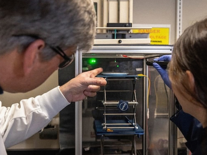
Rheumatologists from the Vasculitis Program at University of Michigan Health have implemented a new method of diagnosing giant cell arteritis, using ultrasound technology.
Ultrasound imaging allows for a non-invasive, faster and less expensive tool to help aid in the diagnosis of giant cell arteritis.
The ultrasound technology looks at the arteries in the head, neck and upper arms for superficial blood vessels that have been affected by the disease.
Testing has shown a high specificity rate, between 92% to 98%, when it comes to detecting giant cell arteritis in patients.
“Giant cell arteritis can be a challenging diagnosis to make. Clinicians have welcomed an additional diagnostic tool in our toolkit. Patients have been excited to have the opportunity to pursue ultrasound for diagnosis and potentially avoid a biopsy,” said Julia Ford, M.D., a clinical assistant professor of rheumatology at U-M Health who pioneered bringing the ultrasound technology to U-M Health.
“Bringing temporal artery ultrasound to U-M Health for the diagnosis of giant cell arteritis has been a great example of teamwork at Michigan Medicine, between the Diagnostic vascular Unit and the Vasculitis Program within the division of rheumatology.”
Learning the ultrasound approach
The technology made its first appearance at University of Michigan Health in December 2022.
Since then, three registered vascular sonographers have been sent to Europe to be trained on the equipment.
“This technology has been used in Europe for a few years with success. My team and I felt that this was something that could benefit our patients by helping them avoid the scheduling, waiting and recovery surrounding a biopsy,” said Andrea Obi, M.D., an assistant professor of vascular surgery at University of Michigan Health.
“The vasculitis program is excited to be able to increasingly offer this technology to our patients to continue improving the care that we give them.”
Since the signs of giant cell arteritis are subtle, an accurate diagnosis requires both a high level of skill from the sonographer and specialized high frequency ultrasound probes that offer exceptional detail of the vascular architecture.
The sonographers are trained to use the ultrasound equipment to measure the inner lining of the blood vessel to assess for inflammation and edema.
This involves very lightly performing the ultrasound to view the vessels in their native configuration as well as using compression to evaluate the blood vessel appearance.
Within 2024, around 182 tests using this technology were performed on patients to diagnose giant cell arteritis.
“On an average, we will get one or two cases a day,” said Ford.
“These cases come from other areas despite rheumatology, such as ophthalmology, neurology and the emergency department.”
Since the technology’s roll out at U-M Health, most patients have been receiving a biopsy in addition to the ultrasound as part of quality assurance.
As familiarity and confidence with this technology improves and more sonographers are trained, the team hopes to begin limiting the need for biopsies in addition to the ultrasounds.
Funding/disclosures: Funding for both the training and new equipment came from the MiAorta program led by an initiative from Jon Eliason, M.D., a professor of vascular surgery at U-M Health and Sandy Brown, U-M Health Diagnostic Vascular Unit Technical Director.
Sign up for Health Lab newsletters today. Get medical tips from top experts and learn about new scientific discoveries every week.
Sign up for the Health Lab Podcast. Add us wherever you listen to your favorite shows.

Health Lab
Explore a variety of health care news & stories by visiting the Health Lab home page for more articles.
Media Contact

Public Relations
Department of Communication at Michigan Medicine
In This Story

Julia A Ford, MD
Clinical Assistant Professor
Stay Informed
Want top health & research news weekly? Sign up for Health Lab’s newsletters today!
Featured News & Stories

Rx Kids linked to reductions in preterm births and low birthweights, fewer NICU admissions

Could preeclampsia become a thing of the past?
Vascular STING activation facilitates natural killer cell anti-tumor immunity in small cell lung cancer

Researchers identify a potential “Achilles heel” of psoriasis
Nanoparticles genetically modify several human cell types