Known as histotripsy, the device not only kills cancer cells but also stimulates the immune system as an added defense.
5:00 AM
Author |
Noninvasive sound technology developed at the University of Michigan breaks down liver tumors in rats, kills cancer cells and spurs the immune system to prevent further spread.
By destroying only 50% to 75% of liver tumor volume, the rats' immune systems were able to clear away the rest, with no evidence of recurrence or metastases in more than 80% of animals.
"Even if we don't target the entire tumor, we can still cause the tumor to regress and also reduce the risk of future metastasis," said Zhen Xu, Ph.D., professor of biomedical engineering at U-M and senior author of the study.
MORE FROM THE LAB: Subscribe to our weekly newsletter
Results also showed the treatment stimulated the rats' immune responses, possibly contributing to the eventual regression of the untargeted portion of the tumor and preventing further spread of the cancer.
The treatment, called histotripsy, noninvasively focuses ultrasound waves to mechanically destroy target tissue with millimeter precision. The relatively new technique is currently being used in a human liver cancer trial in the United States and Europe.
In many clinical situations, the entirety of a cancerous tumor cannot be targeted directly in treatments for reasons that include the mass' size, location or stage. To investigate the effects of partially destroying tumors with sound, this latest study targeted only a portion of each mass, leaving behind a viable intact tumor. It also allowed the team, including researchers at Michigan Medicine and the Ann Arbor VA Hospital, to show the approach's effectiveness under less-than-optimal conditions.
SEE ALSO: Ultrasound technology developed at U-M now clinical trials for liver cancer
"Histotripsy is a promising option that can overcome the limitations of currently available ablation modalities and provide safe and effective noninvasive liver tumor ablation," said Tejaswi Worlikar, M.S., a doctoral student in biomedical engineering. "We hope that our learnings from this study will motivate future preclinical and clinical histotripsy investigations toward the ultimate goal of clinical adoption of histotripsy treatment for liver cancer patients."
Liver cancer ranks among the top 10 causes of cancer related deaths worldwide and in the U.S. Even with multiple treatment options, the prognosis remains poor with five-year survival rates less than 18% in the United States. The high prevalence of tumor recurrence and metastasis after initial treatment highlights the clinical need for improving outcomes of liver cancer.
Even if we don't target the entire tumor, we can still cause the tumor to regress and also reduce the risk of future metastasis.Zhen Xu, Ph.D.
Where a typical ultrasound uses sound waves to produce images of the body's interior, U-M engineers have pioneered the use of those waves for treatment. And their technique works without the harmful side effects of current approaches such as radiation and chemotherapy.
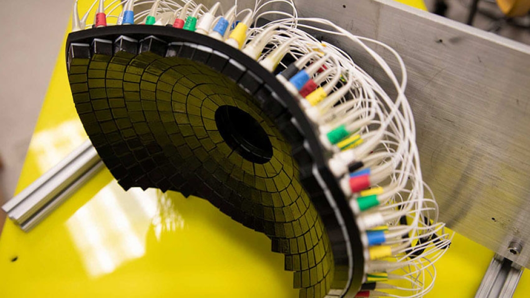
"Our transducer, designed and built at U-M, delivers high amplitude microsecond-length ultrasound pulses—acoustic cavitation—to focus on the tumor specifically to break it up," Xu said. "Traditional ultrasound devices use lower amplitude pulses for imaging."
The microsecond long pulses from UM's transducer generate microbubbles within the targeted tissues—bubbles that rapidly expand and collapse. These violent but extremely localized mechanical stresses kill cancer cells and break up the tumor's structure.
SEE ALSO: New ultrasonic therapy obliterates tissue without physical contact
Since 2001, Xu's laboratory at U-M has pioneered the use of histotripsy in the fight against cancer, leading to the clinical trial #HOPE4LIVER sponsored by HistoSonics, a U-M spinoff company. More recently, the group's research has produced promising results on histotripsy treatment of brain therapy and immunotherapy.
The study was supported by grants from the National Institutes of Health, Focused Ultrasound Foundation, VA Merit Review, U-M's Forbes Institute for Discovery and Michigan Medicine-Peking University Health Sciences Center Joint Institute for Translational and Clinical Research.
U-M retains a financial interest in HistoSonics, as do a number of researchers who were involved in this project and who helped develop the technology, including Xu, who is a company founder, stockholder and consultant. Each stands to benefit financially from the success of the platform.
This story originally appeared on Michigan News.
Paper cited: "Impact of Histotripsy on Development of Intrahepatic Metastases in a Rodent Liver Tumor Model," Cancers. DOI: 10.3390/cancers14071612
Like Podcasts? Add the Michigan Medicine News Break on iTunes or anywhere you listen to podcasts.

Explore a variety of health care news & stories by visiting the Health Lab home page for more articles.
Department of Communication at Michigan Medicine
Want top health & research news weekly? Sign up for Health Lab’s newsletters today!