Researchers Hope to Improve the Masquelet Technique
Orthopaedic surgeons sometimes use the procedure for patients missing a portion of bone. U-M researchers are working on ways to improve the technique.
8:14 AM
Author |
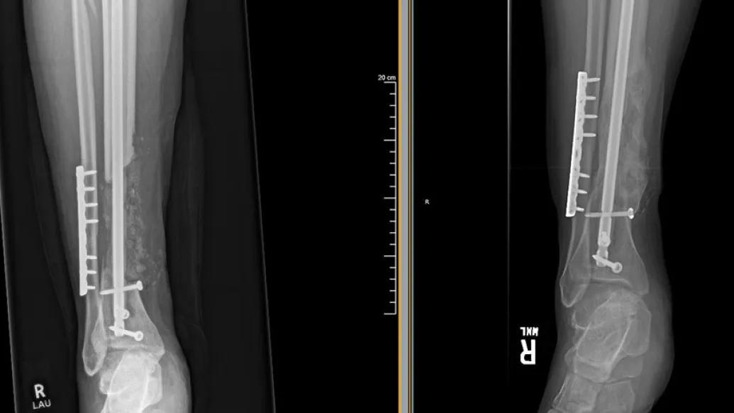
Typically, when a patient experiences a broken bone, an orthopaedic surgeon can realign the broken pieces and stabilize them with implants that allow the bone to heal.
But for some patients who experience a traumatic injury or are born with a sizable bone defect, this method won't work.
"Perhaps the bone has been crushed in a car accident, or the bone is damaged to the point that it won't survive and it's better to remove it," says Mark Hake, M.D., assistant professor of orthopaedic surgery at Michigan Medicine. "In these instances where large pieces of bone are missing, we have to somehow get bone to grow there."
To help these patients, Hake uses the Masquelet technique.
During surgery with the Masquelet technique, orthopaedic trauma surgeons mold a cement spacer where the bone used to be. This allows a bioactive membrane to form around the spacer.
"We leave the spacer in place for six weeks or so and the body forms a layer of tissue around the cement," Hake says. "This tissue forms small blood vessels and secretes proteins that promote bone formation."
LISTEN UP: Add the new Michigan Medicine News Break to your Alexa-enabled device, or subscribe to our daily audio updates on iTunes, Google Play and Stitcher.
After the six weeks are up, the surgical team goes back in for a second surgery.
"We take the spacer out and replace with bone graft taken from another part of the body," Hake says. "The two-stage surgery allows the bone graft to consolidate and form strong, structurally sound bone. If these bone defects don't eventually heal, the patient likely will end up needing an amputation."
We want to eliminate the time period where the spacer has to be left in. We're currently performing the surgery on rats, examining the membrane that forms and looking at what genes are expressed to see if we can come up with a single surgery solution.– Mark Hake, M.D.
Hake notes that while the surgery is not needed in the treatment of the vast majority of fractures, it's important for those with severe injuries.
"For patients that have really terrible injuries, this surgical technique allows us to selvage their limbs and avoid amputation," he says.
Research to better understand and improve the technique
While the Masquelet technique generally has good results, Hake says that research he and colleague, Andrea Alford, Ph.D., research assistant professor of orthopaedic surgery at Michigan Medicine, are working on could build upon the technique and improve it.
"It's interesting because while this two-stage process works, nobody really understands exactly why," Hake says. "We're exploring this in our research and trying to better understand how the body reacts to this process so that it forms bone. And then hopefully from understanding this, we can get a fuller picture of how this promotes bone healing."
MORE FROM THE LAB: Subscribe to our weekly newsletter
In addition to understanding the basic science behind the technique, Hake and Alford are working to improve the recovery time for patients.
"We're trying to figure out how to get the same results, but only doing a single surgery for patients," Hake says. "We want to eliminate the time period where the spacer has to be left in. We're currently performing this surgery on rats, examining the membrane that forms and looking at what genes are expressed to see if we can come up with a single surgery solution."
Future work
Hake and Alford have previously published research on the technique and hope to have results from their current studies later this year. Based on what these results show, they will plan further studies evaluating the possibility of using synthetic bone grafts. Eventually Hake and Alford hope to be able to engineer a membrane to eliminate the need for the second surgery.
Hakes notes that the ability to clinically care for patients and perform research improves overall care for those experiencing a traumatic bone injury.
"Michigan Medicine is unique in that we're not only performing this surgery for our patients and taking care of them clinically, but we're one of only a few institutions across the country doing the basic science research behind it, too," he says.
"The combination of these two is an added benefit for our patients because we're constantly able to explore ways to improve their health and wellbeing after a traumatic bone injury."

Health Lab
Explore a variety of health care news & stories by visiting the Health Lab home page for more articles.
Media Contact
Public Relations
Department of Communication at Michigan Medicine
Stay Informed
Want top health & research news weekly? Sign up for Health Lab’s newsletters today!
Featured News & Stories
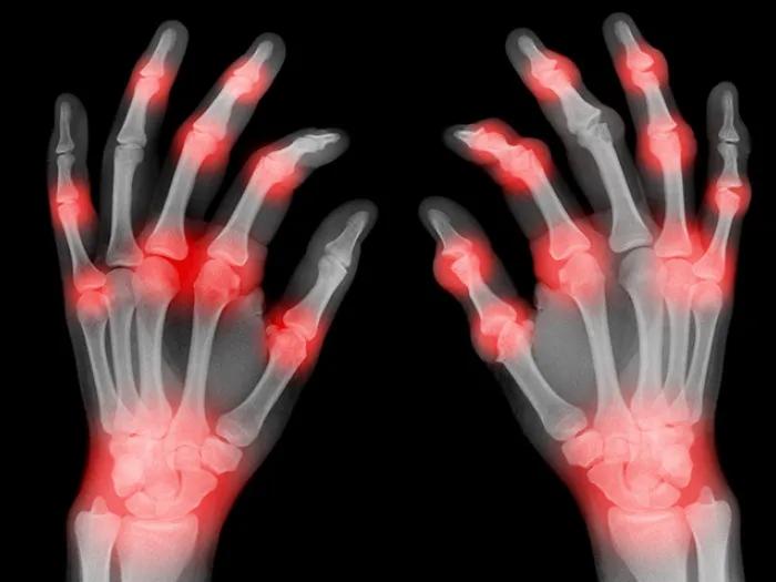
Treating arthritis of the hands

Paying it forward for future Wolverines
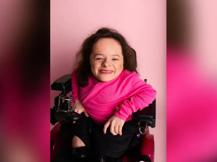
Gift honors reclaimed life and dreams
Two pediatric cancer patients become close friends during treatment
Using a toe for a thumb: Lawn mower accident results in 6-year-old's new digit