A newly developed algorithm shows how a gene is expressed at microscopic resolution
The computational technique makes spatial RNA transcripts more accessible and precise
5:00 AM
Author |

They say a picture is worth a thousand words.
A new method developed by University of Michigan researchers creates images that are worth many gigabytes of data, which could revolutionize the way biologists study gene expression.
Seq-Scope, developed by Jun Hee Lee, Ph.D., Hyun Min Kang, Ph.D., and their colleagues, was first described in the journal Cell in 2021 as the first method to analyze gene expression at sub micrometer-scale spatial resolution.
To compare, a single human hair ranges from 20 to 200 micrometers in width.
The team has since improved Seq-Scope, making it more versatile, scalable, and accessible, which was just published in Nature Protocols.
Additionally, the same group has developed an algorithm for analyzing high-resolution spatial data from Seq-Scope and other technologies, called FICTURE, described in Nature Methods.
“Basically, we are hacking DNA sequencing machines and letting them do all of the hard work,” said Kang, a professor of biostatistics with the U-M School of Public Health.
Researchers use these machines to produce readouts of the transcriptome, the collection of all RNA transcribed from genes with a given cell or tissue.
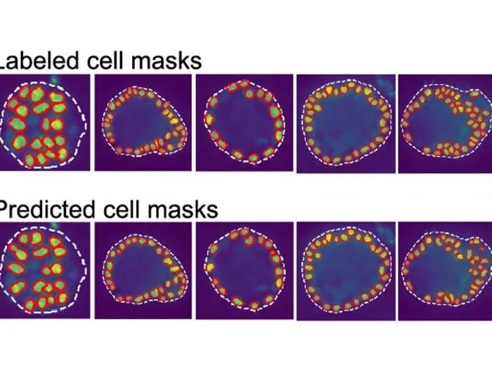
Traditionally, biologists studying genes within a cell or tissue must contend with the fact that a transcriptome has tens of thousands or more genes expressed, too much to make heads or tails of without the help of a computer when it also involves millions of cells.
“The problem is traditionally, there are no computational methods that allow us to understand this data set at microscopic resolution,” said Lee, a professor of Molecular & Integrative Physiology at U-M Medical School.
Lee and Kang’s proof-of-concept method, Seq-Scope demonstrated that a sequencing machine can be repurposed to profile spatially resolved transcriptomes, enabling scientists to see how and where a gene is expressed at microscopic resolution.
The team subsequently has made Seq-Scope even more cost effective, reducing the cost of high resolution spatial transcriptome profiling from upwards of $10,000 to around just $500.
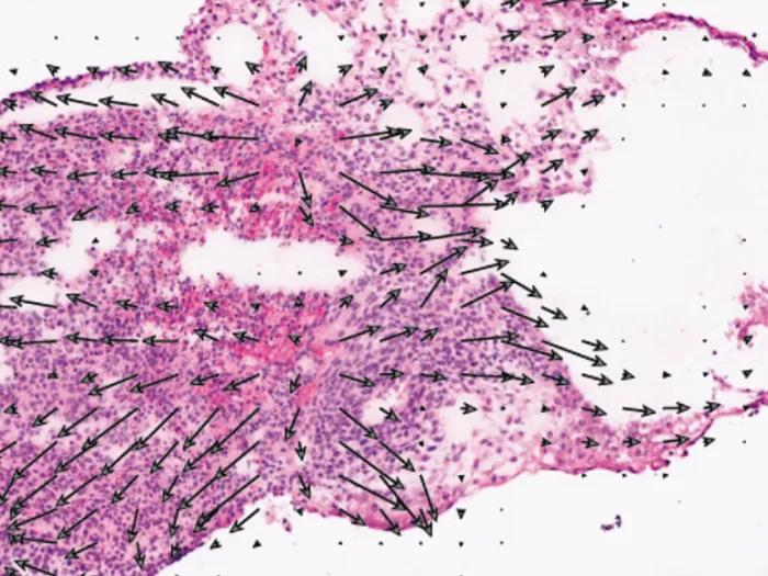
Furthermore, the new FICTURE method enables investigators to analyze massive amounts of data, by pooling the surrounding data together to make a more accurate inference at the micrometer level.
By doing so, they demonstrate, you can see where cell transcripts are located without any bias.
The method generates incredibly detailed images of tissues and cells from its microscopic resolution analysis.
For example, with traditional analysis, “even if you have cell segmentation, if you don’t know exactly which cells are being transcribed and stained, the analysis can be misleading or unclear,” said Kang.
“Using FICTURE, for example, you can see that skeletal muscle tissue from a developing mouse embryo is differentiating into long striated muscle cells from myoblasts.”
“We’re getting a lot of emails from companies and other investigators who previously assumed they wouldn’t be able to do such experiments and analyses. Now they are in the realm of possibility,” said Lee.
U-M’s Advanced Genomics Core co-authored the Seq-Scope protocol paper, contributing by optimizing the use of DNA sequencers.
The AGC is now working to make the Seq-Scope method even more accessible, aiming to disseminate this technology to U-M and the broader scientific community.
"This is exactly the kind of technology we want to bring to as many labs as possible, both here at U-M and beyond," said AGC Director Olivia Koues, Ph.D.
“Our goal is to empower more researchers with cutting-edge spatial transcriptomics capabilities.”
Lee and Kang next hope to develop a way to make the method even more accessible to researchers, enabling them to study genomic expression from beginning to end.
Said Kang, “I think it’s important for computational and experimental investigators to work together to generate new types of data and methods. This is a good example of that type of collaboration.”
Additional authors: Yichen Si, ChangHee Lee, Yongha Hwang, Jeong H. Yun , Weiqiu Cheng, Chun-Seok Cho, Miguel Quiros, Asma Nusrat, Weizhou Zhang, Goo Jun, Sebastian Zöllner, Jun Hee Lee & Hyun Min Kang
Funding/disclosures: Supported in part by National Institutes of Health grants UH3CA268091, R01DK133448, R01AG079163, CA269661, CA290792 and CA260239.
Tech transfer(s)/Conflict(s) of interest: J.H.L. is an inventor on a patent and pending patent applications related to Seq-Scope
Michigan Research Core(s): Advanced Genomics Core
Papers cited:
“Seq-Scope Protocol: Repurposing Illumina Sequencing Flow Cells for High-Resolution Spatial Transcriptomics,” Nature Protocols. DOI: 10.1038/s41596-024-01065-0
“FICTURE: Scalable segmentation-free analysis of submicron resolution spatial transcriptomics,” Nature Methods. DOI: 10.1038/s41592-024-02415-2
Sign up for Health Lab newsletters today. Get medical tips from top experts and learn about new scientific discoveries every week.
Sign up for the Health Lab Podcast. Add us wherever you listen to your favorite shows.

Health Lab
Explore a variety of health care news & stories by visiting the Health Lab home page for more articles.
Media Contact

Public Relations
Department of Communication at Michigan Medicine
In This Story

Jun Hee H Lee
Professor
Stay Informed
Want top health & research news weekly? Sign up for Health Lab’s newsletters today!
Featured News & Stories

Studying neurons using neurons

How AI is helping emergency physicians learn from their patients

The algorithm will see you now? Patients say not without a doctor nearby

AI tool can predict heart failure from genetic and health record data
AI reveals hidden features of a developing embryo model