Nanoscale images of a blood clotting protein complex reveal a secret to the clotting chain reaction
The findings help solve a decades-old puzzle in biological chemistry
5:00 AM
Author |

If you’ve ever accidentally sliced yourself on a broken glass or a piece of paper, you may have noticed that the bleeding can be hard to stop.
Scientists have long wondered how the cascade of events that leads to blood clotting is triggered, especially since the process has life and death consequences.
Too little clotting and you bleed out, while too much can cause deadly clots leading to a heart attack or stroke.
New detailed 3D structures of blood clotting proteins, made possible by using cryogenic-electron microscopy (Cryo-EM), have solved a mystery vexing biological chemists for more than 30 years.
Cryo-EM is a structural approach where biological samples are trapped in a layer of non-crystalline ice and imaged using powerful electron microscopes.
The U-M Life Sciences Institute, with support of the U-M Bioscience Initiative and Arnold and Mabel Beckman Foundation, has established a state-of-the art cryo-EM facility that made the research possible.
James Morrissey, Ph.D., professor of biochemistry at U-M Medical School, has been studying the various proteins involved in clotting since the 1980s.
To solve this structure, he teamed up with cryo-EM expert, Melanie Ohi, Ph.D., a professor of cell and developmental biology at U-M Medical School and research professor in the U-M Life Sciences Institute.
“Most of the blood clotting proteins are soluble proteins that circulate in your blood, and there’s one missing protein that is housed on the surface of cells outside of the vasculature,” he explained.
The normal clotting process involves an enzyme with two sub-units, tissue factor and factor VIIa. When that combo links up on a cell surface, it kicks off the clotting cascade, he says.
The cell membrane plays an important role in whether clotting will happen.
“A happy, healthy resting cell won’t bind these blood clotting proteins,” he said. But an injury causes the particular phospholipids from the lipid bilayer that makes up the cell membrane to flip to the outside of the cell, where the blood clotting proteins bind to them, he explained.
In the case of a profusely bleeding paper cut, “it could be that if you don’t damage enough cells and don’t expose enough of these phospholipids of the surface of the damaged cells to recruit blood clotting proteins, then clotting is slowed,” Morrissey noted.
This process has been exceedingly hard to study so Morrissey and his colleagues used cryo-EM to determine 3D structures that allowed them to build an atomic model of the protein interactions when associated with a lipid nanodisc.
They found that the tissue factor/factor VIIa complex binds to the second protein in the clotting cascade, called factor X by moving a small site on tissue factor out of the way, changing the structure so that factor X can dock onto that site, like two puzzle pieces fitting together (see movie).
“It was exciting to determine a structure that helps solve the long-standing mystery for why tissue factor can activate the blood clotting cascade only when specific lipids are found on the outside of a cell. For me this work highlights two strengths of doing research at the University of Michigan – the ease at establishing successful cross-disciplinary collaborations and access to one of the best cryo-electron microscopy facilities in the world. Working in this type of environment makes it fun to tackle challenging structural questions,” said Ohi.
Their results are published in a recent issue of the American Society of Hematology journal, Blood.
The findings help explain “something that people have known about for years: that this part of tissue factor was important in recognizing the substrate and allowing the reaction to go. But nobody came up with how the proteins actually dock together,” said Morrissey.
Anticoagulant drugs like Warfarin, prescribed to treat dangerous blood clotting, work by weakening the ability of these proteins to interact at the cell membrane, but they worked without scientists fully understanding what was happening.
This basic science work finally provides new insights into the mechanism behind it.
Additional authors: Josepha C Sedzro, Amanda L. Photenhauer, Fabienne Birkle, Katarina Meze, Alex Mortenson, Cade Duckworth, Po-Chao Wen, Sarah Kearns, Michael A. Cianfrocco, Emad Tajkhorshid, Melanie D. Ohi
Paper cited: “Cryo-EM structure of the tissue factor/factor VIIa complex with a factor X mimetic reveals a novel allosteric mechanism,” Blood. DOI: 10.1182/blood.2025029430
Sign up for Health Lab newsletters today. Get medical tips from top experts and learn about new scientific discoveries every week.
Sign up for the Health Lab Podcast. Add us wherever you listen to your favorite shows.

Health Lab
Explore a variety of health care news & stories by visiting the Health Lab home page for more articles.
Media Contact
Public Relations
Department of Communication at Michigan Medicine
In This Story
James H Morrissey
Professor
Melanie D Ohi PhD
Research Professor
Stay Informed
Want top health & research news weekly? Sign up for Health Lab’s newsletters today!
Featured News & Stories
Studying neurons using neurons
This “essential” nutrient likely has no health benefit
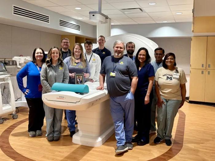
New prostate biopsy technique shows potential future of MRI

An online intervention can help cancer patients share genetic testing results with family

The algorithm will see you now? Patients say not without a doctor nearby