Improved model system allows researchers to study embryo development
Their study uncovers more about how embryos are formed
5:00 AM
Author |
Research from Ph.D. candidate Bohan Chen in the lab of Idse Heemskerk of the Department of Cell and Developmental Biology at the University of Michigan Medical School and their colleagues improves upon a popular experimental model and in doing so, reveals more of the inner workings of a critical period during the formation of an embryo.
Scientists study development in part to understand what can go wrong as the body’s structures are formed.
The ultimate hope is to be able to prevent birth defects and identify causes of failed pregnancy.
However, studying embryos in a laboratory is fraught with important ethical and technical considerations.
To alleviate these concerns, many researchers turn to simple 2D structures made up of stem cells called gastruloids.
Gastruloids are grown, or cultured, in vitro (in a dish) and model some of the earliest moments of development without the potential to develop into a human being.
Gastruloids recreate aspects of a process called gastrulation in experiments.
During gastrulation, a so-called primitive streak of cells gives rise to the three embryonic layers of the cells that ultimately make up the body plan: the ectoderm (outer layer), which gives rise to the skin, nervous system and other outer structures; the mesoderm (middle layer), which becomes the heart, muscles, bones and other internal structures; and the endoderm (the inner layer) that becomes the GI tract, lungs, liver and other organs.
However, the in vitro culturing of gastruloids has typically only been sustained for up to two days, where after, the cells became disorganized and stop developing.
“We started using different medium and made several other improvements in the model,” said Heemskerk.
“And when we tried growing the cells for more than two days, it actually worked and did some really interesting things.”
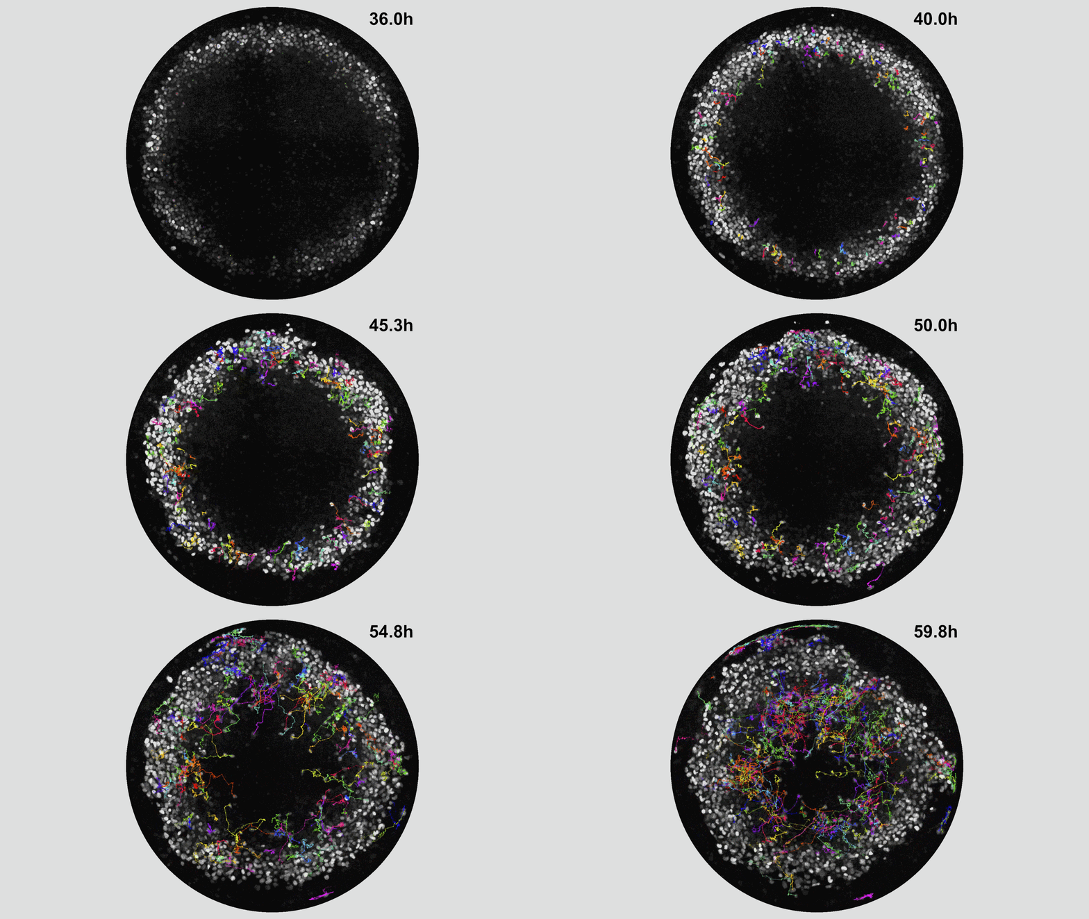
For starters, the team saw that the stem cells of the developing mesoderm began to travel underneath the original single layer of cells, forming a multi-layered structure just as it would in a real embryo.
“This process is very hard to visualize; we don’t even know how it works in mice,” said Heemskerk.
Yet, their extended model enabled them to see that the mesoderm stem cells’ migration is directed from the edge of the group of cells to the center.
“That means there’s something—we don’t yet know what—telling them which way to go. We now have a setup in which we can figure out what is guiding their movement,” said Heemskerk.
This is critically important for figuring out what goes wrong in certain cases, such as with the generation of a congenital heart defect.
The team also determined that the mesoderm cells in a gastruloid come in several different subtypes that express different genes, which they visualized using fluorescence.
“By looking at what gene the cell is expressing you can kind of see which organ it will ultimately become,” he said.
This begs the question, notes Heemskerk, do cells know what their fate is prior to migration or does where they end up determine what they will be?
Heemskerk hopes to continue this work using their extended model to answer these and more questions about mammalian development.
“This is a simple model that allows us to see things that would be very difficult in a complex 3D structure but that also captures biological phenomena while allowing us to avoid concerns that come with working with embryos.”
Paper cited: “Extended culture of 2D gastruloids to model human mesoderm development,” Nature Methods. DOI: 10.1038/s41592-025-02669-4

Health Lab
Explore thousands of health news & research stories by visiting the Health Lab homepage for more.
Media Contact

Public Relations
Department of Communication at Michigan Medicine
In This Story
Idse Heemskerk, PhD
Associate Professor
Stay Informed
Want top health & research news weekly? Sign up for Health Lab’s newsletters today!
Featured News & Stories
Comprehensive DNA sequencing reveals hidden drivers of cancer

Many older adults make Medicare decisions quickly and most don’t change plans, poll finds
‘Molecular glue’ stabilizes protein that inhibits development of non-small cell lung cancer and uterine cancer

What’s the latest on 7-OH and kratom availability and addiction care?

Depression in OBGYN residents needs more attention, study suggests