By hijacking the function of a key element of the cell, the coronavirus gains ground
10:59 AM
Author |
In March 2020, as laboratories around the University of Michigan Medical School were shutting down operations as part of the COVID-19 public health emergency, scientists who were already equipped to study viruses looked for ways they could help stop the pandemic.
A recent paper from the lab of Billy Tsai, Ph.D., of the Department of Cell & Developmental Biology details their contribution to the cause by way of a highly complicated experiment designed to reveal how SARS-CoV-2 causes infection by hijacking cellular machinery.
“This was a new virus to our lab and there were a lot of barriers there,” commented Jeffrey Williams, a graduate student who performed many of live virus experiments. It sounds easy because our lab works with a bunch of viruses—such as dengue, Zika and SV40—but each virus does different things and replicates in different ways.”
As part of their pivot to studying SARS-CoV-2, the team had to familiarize themselves with coronavirus biology. Then letting the literature guide them, they turned to the wealth of information being churned out about SARS-CoV-2 genetics by researchers around the world.
“There were a couple of big proteomics papers that came out showing how proteins of the virus interacted with proteins in host cells that we referenced,” said Williams.
Because the lab had previous work studying proteins associated with a cellular organelle called the endoplasmic reticulum, they started there. The ER, known for its highly convoluted shape, produces proteins for the cell to function, among other tasks.
“A lot of viruses have to go to the ER in order to cause infection,” said Yu-Jie Jay Chen, Ph.D., a research investigator on the study. Screening several of the proteins in the ER, they discovered that if they blocked certain proteins called reticulons inside cells, SARS-CoV-2 infection was reduced.
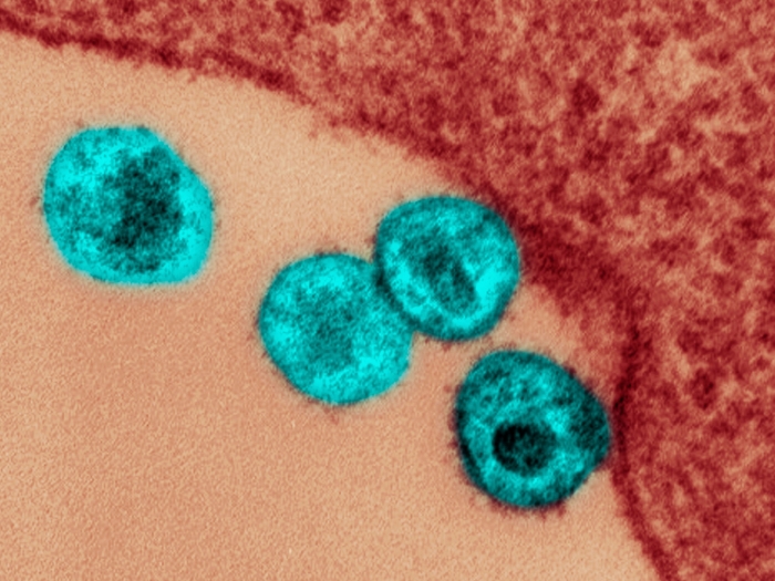
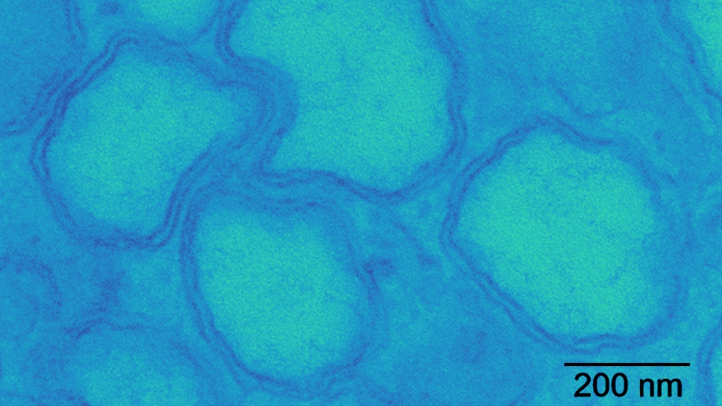
Reticulons are morphogenic proteins, and they give the ER its shape, explained Chen. “We discovered that SARS-CoV-2 uses these proteins to generate a special organelle called a double membrane vesicle.”
The double membrane vesicle is a pod-like structure in which the virus makes copies of itself while hiding from the immune system of the host. This was true across several SARS-CoV-2 variants, they add.
Could blocking reticulon function be key to stopping SARS-CoV-2? Perhaps not, note the researchers.
“The danger is obviously you’d be mucking around with basic cellular function,” said Tsai. “But perhaps you could bring the reticulon level down just enough to support normal biology of the cell but block infections.”
The team plans to continue to study ER biology to identify additional proteins important to the lifecycle of SARS-CoV-2.
Additional authors on this paper include Woo Jung Cho and Andrew W. Tai.
Tsai is funded by National Institutes of Health grant, RO1 AI170514.
Paper cited: “Reticulons promote formation of ER-derived double-membrane vesicles that facilitate SARS-CoV-2 replication,” Journal of Cell Biology. DOI: 10.1083/jcb.202203060

Explore a variety of healthcare news & stories by visiting the Health Lab home page for more articles.
Department of Communication at Michigan Medicine
Want top health & research news weekly? Sign up for Health Lab’s newsletters today!