Early intervention and expertise crucial for treating pediatric craniofacial disorder
A pediatric neurosurgeon weighs in on treatment options for craniosynostosis
5:00 AM
Author |
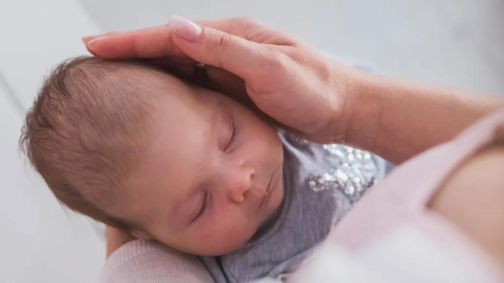
Approximately one in 2,000 babies is born with craniosynostosis, a type of craniofacial disorder in which a baby's skull fuses too soon.
This birth disorder causes the fibrous joints between the bones of a baby’s skull to close before the brain is fully developed, leading to an irregular head shape.
As the child matures, this early fusion of the skull can lead to vision issues, headaches, increased pressure on the brain and neurocognitive delays.
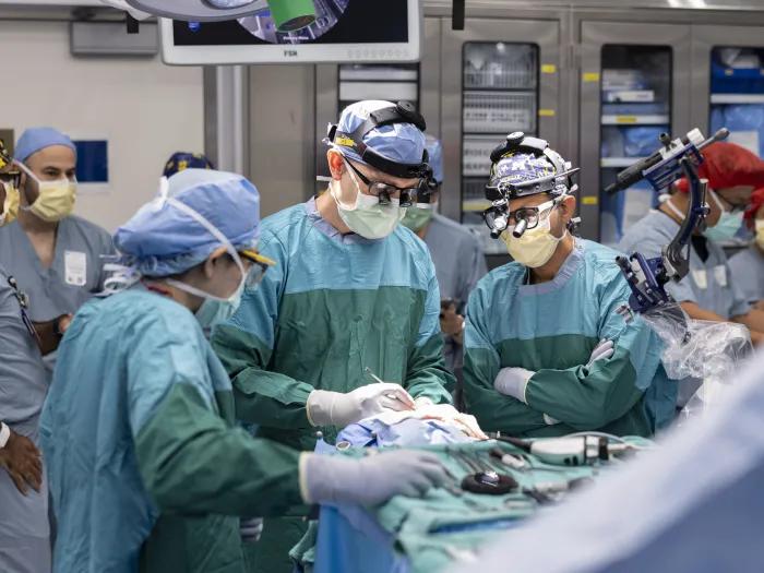
Pediatric neurosurgeon Suresh Magge, M.D., clinical professor of neurosurgery at University of Michigan Health C.S. Mott Children’s Hospital and a nationally recognized leader in the surgical management of craniosynostosis, answers important questions about the condition, stressing the critical need for early intervention.
How does craniosynostosis present in a newborn?
A newborn child’s skull is made of multiple skull plates designed to remain open at the junctions early in life, allowing the brain to grow symmetrically.
When a child is born with craniosynostosis, one or more of those junctions, or sutures, fuses too soon, resulting in an abnormal head shape that can restrict skull growth.
As the child grows, this abnormal head shape can not only affect his or her psychosocial well-being but can also cause increased intracranial pressure, or increased pressure on the brain.
What steps should your pediatrician take when a newborn is suspected of having craniosynostosis?
A pediatrician should recommend parents have the newborn evaluated at a center that offers a highly skilled multidisciplinary pediatric team of craniofacial experts.
The most important thing is that the infant be seen as soon as possible as successful treatment for craniosynostosis depends on early referral and diagnosis.
What should you look for when deciding where to seek care for your child?
This condition requires a team of pediatric experts who are able to offer the most advanced treatment options to ensure the best possible outcome.
It's important that the center offers both open and minimally invasive surgical options for craniosynostosis, provided by a team that specializes in the condition.
At C.S. Mott Children’s Hospital, we have a multidisciplinary team with highly trained and highly experienced experts to treat craniosynostosis.
The team includes some of the most experienced pediatric neurosurgeons and pediatric plastic surgeons, and we offer both minimally invasive and open surgical options.
The team also includes dentistry, maxillofacial surgery and social work to provide long term follow up care, with each child followed throughout childhood.
Our craniofacial program has all the necessary pediatric specialists ready to evaluate the child, propose appropriate treatment options and provide the best treatment using the least invasive methods.
We offer expertise in both open surgical correction and minimally invasive endoscopic surgery, with an emphasis on finding the best treatment for each child.
As a neurosurgeon, how do you determine the best treatment plan?
After a thorough clinical exam by the craniofacial team, a CT scan may also be done to evaluate the anatomy of the skull.
Based on this information, our specialists determine if a child has craniosynostosis and the best treatment option.
There are two main types of treatment: open surgical repair of craniosynostosis and minimally invasive/endoscopic surgery.
Open surgical repair may also be known as calvarial vault reconstruction or fronto-orbital advancement, depending on the specific sutures fused.
In this surgery, an incision is made from ear to ear, and then the surgical team, composed of a pediatric neurosurgeon and pediatric plastic surgeon, makes cuts in the skull and then reconstructs the cranial vault to normalize the skull shape and allow more room for the brain.
This type of procedure is typically done between the ages of 6 to 12 months.
Minimally invasive/endoscopic surgery is a less invasive surgery, in which one or two small incisions are made in the scalp.
The fused portion of the skull is removed with the assistance of an endoscope, or small camera.
This surgery generally involves smaller incisions, shorter surgery, shorter hospital stays and less blood loss (with lower chance of blood transfusion than open surgery).
The surgery is done at an earlier age than open surgery, usually before 3 or 4 months of age for optimal outcomes.
After surgery, the child would wear a cranial molding helmet to gradually correct the skull shape over a few months.
We have excellent outcomes with minimally invasive/endoscopic surgery as well as open surgery, but if diagnosed early, a child can be a candidate for the less invasive surgery.
This is why early referral to our center is very important if the pediatrician suspects craniosynostosis.
What are the latest innovations in treating craniofacial disorder?
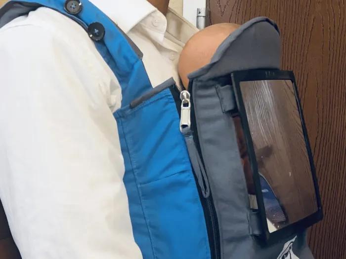
In addition to special expertise in minimally invasive endoscopic repair, C.S. Mott Children’s Hospital is also one of only a few children's hospitals in the country to have a state-of-the-art three-dimensional camera system installed in the craniofacial clinic.
The camera allows us to get highly accurate photos before and after surgery to track a child’s progress.
What developments will make the biggest impact on the treatment of craniofacial disorder?
We continue to improve our techniques and make surgery less invasive.
Forty years ago, the only option was a very invasive open surgery, and now we have the ability correct the disorder through very small incisions and in a much less invasive manner than ever before.
Both surgeries can lead to excellent outcomes, so we find an individualized treatment plan that is tailored for each child.
It's important that parents have all options at their disposal to have the best treatment for a child diagnosed with craniosynostosis.
Sign up for Health Lab newsletters today. Get medical tips from top experts and learn about new scientific discoveries every week.
Sign up for the Health Lab Podcast. Add us wherever you listen to your favorite shows.

Health Lab
Explore a variety of health care news & stories by visiting the Health Lab home page for more articles.
Media Contact
Public Relations
Department of Communication at Michigan Medicine
In This Story
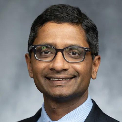
Suresh N Magge, MD
Clinical Professor
Stay Informed
Want top health & research news weekly? Sign up for Health Lab’s newsletters today!
Featured News & Stories
Expert complex heart surgery team saves baby with rare genetic heart condition
Medical student’s invention aims to help infants with jaundice
Research may help better predict outcomes in kids with congenital cytomegalovirus
12-year-old shares journey with sickle cell anemia on Capitol Hill
University of Michigan implants first-in-human Paradromics wireless brain-computer interface, designed to restore communication