‘Concerning’ CT scans may cause unnecessary hospitalization for some pulmonary embolism patients
Imaging results could represent an important barrier to discharge of low risk patients, researchers say
5:00 AM
Author |
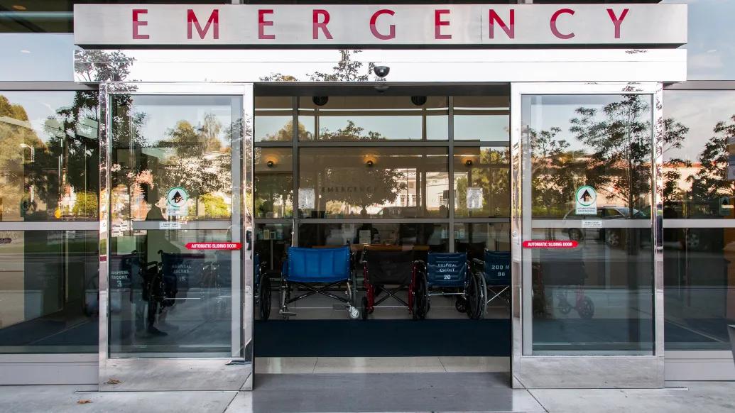
Of approximately 250,000 Americans diagnosed with acute pulmonary embolism, or PE, in emergency departments each year, most are hospitalized.
But Michigan Medicine research, published in JAMA Network Open, finds that some patients with PE, a blood clot in one or more pulmonary arteries, may be hospitalized unnecessarily due to computed tomography, or CT, imaging results rather than clinical risk factors.
Approximately 40% of the patients in the study had low risk pulmonary embolism, as defined by the Pulmonary Embolism Severity Index, or PESI score. Roughly half of the low risk patients had CT imaging features that physicians consider “concerning”, and these patients fared just as well in the hospital as those whose CT scans showed no concerning findings.
“The results of this study are going to come as a surprise to most emergency physicians, who have been taught for years to regard large, centrally located, or “saddle”, PEs as true medical emergencies,” said senior author Colin Greineder, M.D., Ph.D., an assistant professor of emergency medicine and pharmacology at University of Michigan Medical School.
“In fact, our findings suggest that CT findings alone might not confer as much risk as we thought. In our population, if the patient’s pulse, rate of breathing, oxygen level and other clinical factors indicated low risk, then ‘concerning’ CT findings by themselves were not associated with worse clinical outcomes.”
Researchers assessed data from more than 800 patients at U-M Health diagnosed with acute pulmonary embolism in the adult emergency department between late 2016 and the end of 2019.
Using the PESI score, patients were divided into low and high risk groups, with the low risk group further divided based on the presence of concerning CT findings. These included large and centrally located PEs, blood clots on both sides of the lung, evidence of strain on the heart due to the blockage and associated infarction, or death, of a region of lung tissue.
Our report suggests that these imaging findings may be an important barrier to safe discharge."
Geoffrey Barnes, M.D., M.Sc.
Within a month of coming to the emergency department, 18% of patients with high risk PESI scores died, whereas none of the low risk patients with concerning CT imaging findings died. These patients also rarely required intensive care, and their lengths of stay in the hospital were no different than low risk patients without ‘concerning’ CT imaging findings.
“Clinical outcomes were similar, but we found plenty of evidence that emergency physicians are managing these patients differently,” Greineder said.
“The rate of discharge and home management was about four times lower in low risk patients with concerning CT imaging findings, and they were more likely to have bedside ultrasounds performed in the emergency department and echocardiograms while in the hospital.”
The study also investigated how often emergency physicians activated the PE Response Team, a multi-disciplinary group designed to make rapid, shared decisions on the management of patients with intermediate and high risk PE at U-M Health.
Response team activation was almost four times more likely in low risk patients with concerning CT imaging findings despite their low risk PESI scores.
SEE ALSO: Complications for procedure to open clogged pulmonary arteries decrease significantly
Over the past two decades, a strong evidence base has been established to support safe outpatient management of pulmonary embolism, but adoption by emergency medicine clinicians has been slow and remains limited, says co-author Geoffrey Barnes, M.D., M.Sc., associate professor of cardiology-internal medicine at U-M Medical School.
“We have tools like the Pulmonary Embolism Severity Index and biomarkers that allow for appropriate management of these patients, but they do not typically include those ‘concerning’ CT findings. Our report suggests that these imaging findings may be an important barrier to safe discharge.”
The results, researchers say, require further research and confirmation to verify that these findings are replicated in other hospital systems and patient populations.
“Like other groups, we are already working to overcome other barriers to outpatient management of low risk PE,” said first author Connor O’Hare, M.D., an emergency medicine resident at U-M Health. “If our current findings are reproduced in other studies and hospital systems, then we will need effective implementation strategies to overcome the perceived risk associated with ‘concerning’ CT imaging findings in otherwise low risk patients.”
Additional authors include: Kelsey A. Grace, M.D., S. Nabeel Hyder, M.D., Michael Stover, Amber L. Liles, M.D., Minhaj S. Khaja, M.D., James A. Cranford, Ph.D., Keith E. Kocher, M.D., all of University of Michigan, and William J. Schaeffer, D.O., now of Medical College of Wisconsin.
This work was funded by an EM Residency Research Development Grant (RDG) Award from the University of Michigan and the National Institutes of Health (K08-HL130430 and R01-HL163438).
Khaja reported receiving speaking honoraria from Penumbra and Boston Scientific, volunteering for the advisory board of Medtronic, and receiving grants from Boston Scientific outside the submitted work. Kocher reported that grant funding from Blue Cross Blue Shield of Michigan and Blue Care Network supports the Michigan Emergency Department Improvement Collaborative, a quality improvement network. Barnes reported receiving consulting fees from Pfizer, Bristol-Myers Squibb, Janssen Pharmaceuticals, Bayer and Abbott Vascular and receiving consulting fees and serving as a trial site principal investigator for Boston Scientific outside the submitted work. No other disclosures were reported.
Paper cited: “Adverse Clinical Outcomes Among Patients With Acute Low-risk Pulmonary Embolism and Concerning Computed Tomography Imaging Findings,” JAMA Network Open. DOI: 10.1001/jamanetworkopen.2023.11455

Health Lab
Explore thousands of health news & research stories by visiting the Health Lab homepage for more.
Media Contact
Public Relations
Department of Communication at Michigan Medicine
Stay Informed
Want top health & research news weekly? Sign up for Health Lab’s newsletters today!
Featured News & Stories

E-bikes are faster than ever. Are families prepared for the risks?

Scientists discover that bacteria use an understudied polymer found in all life to protect cell functions during stress

Aortic valve replacement: Understanding TAVR and SAVR

The hidden life of parasites like Cyclospora
More than half of U.S. adults now qualify for statins under new guidelines