Artificial intelligence predicts genetics of cancerous brain tumors in under 90 seconds
Researchers hope it will improve diagnosis and treatment, as well as clinical trial enrollment
12:00 PM
Author |
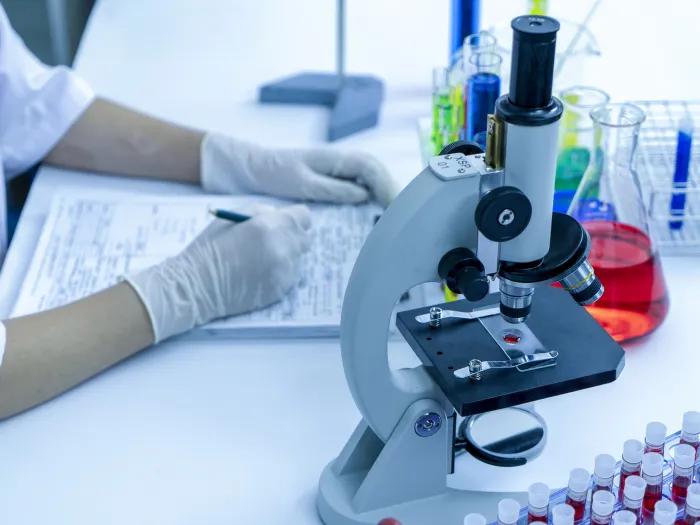
Using artificial intelligence, researchers have discovered how to screen for genetic mutations in cancerous brain tumors in under 90 seconds — and possibly streamline the diagnosis and treatment of gliomas, a study suggests.
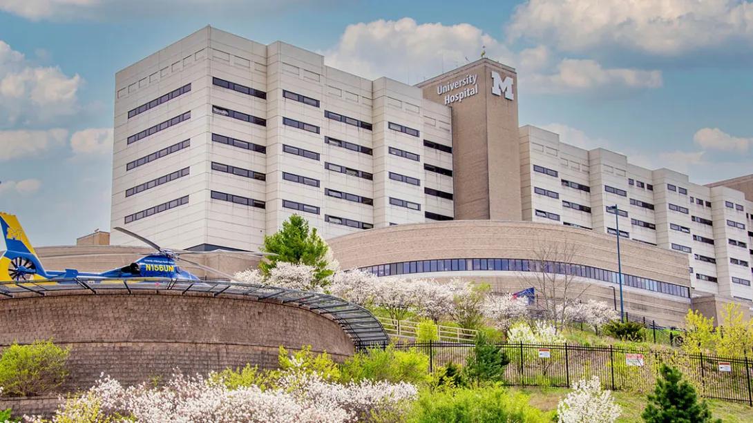
A team of neurosurgeons and engineers at Michigan Medicine, in collaboration with investigators from New York University, University of California, San Francisco and others, developed an AI-based diagnostic screening system called DeepGlioma that uses rapid imaging to analyze tumor specimens taken during an operation and detect genetic mutations more rapidly.
In a study of more than 150 patients with diffuse glioma, the most common and deadly primary brain tumor, the newly developed system identified mutations used by the World Health Organization to define molecular subgroups of the condition with an average accuracy over 90%. The results are published in Nature Medicine.
“This AI-based tool has the potential to improve the access and speed of diagnosis and care of patients with deadly brain tumors,” said lead author and creator of DeepGlioma Todd Hollon, M.D., a neurosurgeon at University of Michigan Health and assistant professor of neurosurgery at U-M Medical School.
Molecular classification is increasingly central to the diagnosis and treatment of gliomas, as the benefits and risks of surgery vary among brain tumor patients depending on their genetic makeup. In fact, patients with a specific type of diffuse glioma called astrocytomas can gain an average of five years with complete tumor removal compared to other diffuse glioma subtypes.
DeepGlioma creates an avenue for accurate and more timely identification that would give providers a better chance to define treatments and predict patient prognosis."
Todd Hollon, M.D.
However, access to molecular testing for diffuse glioma is limited and not uniformly available at centers that treat patients with brain tumors. When it is available, Hollon says, the turnaround time for results can take days, even weeks.
“Barriers to molecular diagnosis can result in suboptimal care for patients with brain tumors, complicating surgical decision-making and selection of chemoradiation regimens,” Hollon said.
Prior to DeepGlioma, surgeons did not have a method to differentiate diffuse gliomas during surgery. An idea that started in 2019, the system combines deep neural networks with an optical imaging method known as stimulated Raman histology, which was also developed at U-M, to image brain tumor tissue in real time.
SEE ALSO: Artificial Intelligence Improves Brain Tumor Diagnosis (michiganmedicine.org)
“DeepGlioma creates an avenue for accurate and more timely identification that would give providers a better chance to define treatments and predict patient prognosis,” Hollon said.
Even with optimal standard-of-care treatment, patients with diffuse glioma face limited treatment options. The median survival time for patients with malignant diffuse gliomas is only 18 months.
While the development of medications to treat the tumors is essential, fewer than 10% of patients with glioma are enrolled in clinical trials, which often limit participation by molecular subgroups. Researchers hope that DeepGlioma can be a catalyst for early trial enrollment.
“Progress in the treatment of the most deadly brain tumors has been limited in the past decades- in part because it has been hard to identify the patients who would benefit most from targeted therapies,” said senior author Daniel Orringer, M.D., an associate professor of neurosurgery and pathology at NYU Grossman School of Medicine, who developed stimulated Raman histology. “Rapid methods for molecular classification hold great promise for rethinking clinical trial design and bringing new therapies to patients.”
Additional authors include Cheng Jiang, Asadur Chowdury, Akhil Kondepudi, Arjun Adapa, Wajd Al-Holou, Jason Heth, Oren Sagher, Maria Castro, Sandra Camelo-Piragua, Honglak Lee, all of University of Michigan, Mustafa Nasir-Moin, John Golfinos, Matija Snuderl, all of New York University, Alexander Aabedi, Pedro Lowenstein, Mitchel Berger, Shawn Hervey-Jumper, all of University of California, San Francisco, Lisa Irina Wadiura, Georg Widhalm, both of Medical University Vienna, Volker Neuschmelting, David Reinecke, Niklas von Spreckelsen, all of University Hospital Cologne, and Christian Freudiger, Invenio Imaging, Inc.
This work was supported by the National Institutes of Health, Cook Family Brain Tumor Research Fund, the Mark Trauner Brain Research Fund, the Zenkel Family Foundation, Ian’s Friends Foundation and the UM Precision Health Investigators Awards grant program.
Paper cited: “Artificial-intelligence-based molecular classification of diffuse gliomas using rapid, label-free optical imaging,” Nature Medicine. DOI: 10.1038/s41591-023-02252-4

Health Lab
Explore thousands of health news & research stories by visiting the Health Lab homepage for more.
Media Contact
Public Relations
Department of Communication at Michigan Medicine
Stay Informed
Want top health & research news weekly? Sign up for Health Lab’s newsletters today!
Featured News & Stories

Many young adults may not be ready to manage their own health care
CRISPR acts as commander-in-chief for backup defenses in bacteria
Scientists discover that bacteria use an understudied polymer found in all life to protect cell functions during stress
The hidden life of parasites like Cyclospora
Long friendship inspires urology resident fund
