Enhancing Eye Care with a Smartphone
Consumers use smartphones for just about everything. A U-M ophthalmologist wants to bring them into the doctor’s office, too, as easily accessible retinal cameras.
7:00 AM
Author |
From photo sharing to driving directions to grocery lists, smartphones are integrated into our everyday lives. Millions of people use them for their own health with wearable devices to track heart rate, sleep quality and steps.
SEE ALSO: How Printing a 3-D Skull Helped Save a New One
A University of Michigan ophthalmologist is working to bring the health applications, plus the convenience and reliability of smartphones, into the doctor's office as well to enrich eye care.
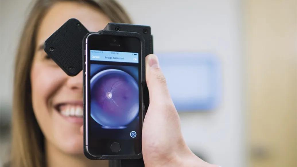
"We want to use an iPhone for its camera, its computational power and its connectivity to turn the smartphone into a retinal camera, which is traditionally a challenging and expensive tool," says Tyson Kim, M.D., Ph.D., a former resident ophthalmologist at U-M's Kellogg Eye Center. Kim is part of a team that came together at the University of California Berkeley (Daniel Fletcher, Ph.D.) and University of California San Francisco (Todd Margolis, M.D., Ph.D., currently Washington University in St. Louis) to invent a device called the CellScope Retina, which they're testing right now.
"The back of the eye is the only place on the body you can see blood vessels and nerves directly," Kim says. "There are a lot of applications, but the exam is rarely performed since it's so inaccessible."
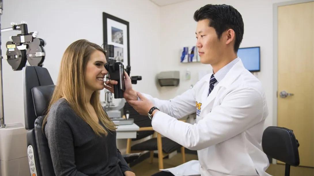
A smartphone slides into the CellScope Retina device to turn it into a functioning retinal camera. Then, a primary care physician or other health care provider could image the back of a patient's eye in the doctor's office. An accompanying app makes it easy to use.
"The goal is to put a device in the hands of PCPs," Kim says. "It simplifies everything so the patient doesn't need to worry about referrals to specialists unless absolutely needed, and the PCPs are able to take better care of patients, improve their outcome metrics and gain a new billable procedure."
Removing barriers
A retinal camera has traditionally cost upward of $100,000, and even though the equipment has become a bit more reasonably priced lately, it is still immobile and hard to use. The professionals who operate retinal cameras in eye clinics traditionally go through two-year training programs.
A simpler, more cost-effective way to take the same images could remove the barriers to widespread exams.
"Because everyone is so familiar with a smartphone already, we enable general health care providers to easily and quickly capture good retinal images," Kim says.
Retinal exams for people with diabetes
People with diabetes in particular should receive annual retinal exams, but many do not go for a checkup until vision problems are advanced, even if their primary care doctor provides a referral. Diabetic retinopathy is a leading cause of vision loss and blindness among adults, according to the National Eye Institute.
SEE ALSO: The Artificial Heart You Take with You
This diabetic side effect can be prevented or delayed with proper exams, and patient's insurance would cover this preventive activity.
"Patients with diabetes who don't know they have the beginnings of vision loss wouldn't think to ask for a referral, but when it's covered as part of the PCP visit the percentage of patients receiving exams should increase," Kim says.
Other applications
Manifestations of systemic disease can often be identified in the back of the eye. If accessibility of retinal imaging were increased, doctors could immediately image the back of the eye to help identify trauma or child abuse, increased intracranial pressure as with a brain tumor, high blood pressure, evidence of embolic heart disease and more.
"A simplified device like this could also be used in low-resource regions of the world to expand eye care to places in need," Kim says. The CellScope Retina is undergoing continued development at the U-M Kellogg Eye Center with the goal of not requiring pharmacological dilation of the patient's eye.
Kim received the Michigan Translational Research and Commercialization Fast-Forward Medical Innovation Award for his work, based on its high potential for translation into the commercial industry. The team is currently performing usability testing, then will begin beta testing in a primary care setting.
Disclosure: Kim is one of the inventors and part of a multi-institutional collaboration developing the CellScope Retina. In addition to Kim, Margolis and Fletcher, other key members of the team include Yannis Paulus, M.D. (U-M) and Maria Woodward, M.D. (U-M).

Health Lab
Explore thousands of health news & research stories by visiting the Health Lab homepage for more.
Media Contact

Public Relations
Department of Communication at Michigan Medicine
Stay Informed
Want top health & research news weekly? Sign up for Health Lab’s newsletters today!
Featured News & Stories
Innovative children’s vision research gets a boost from the Knights Templar Eye Foundation
Many hospitals still don’t do enough to prevent UTIs from catheters

Is the United States Lagging Behind Europe in Memory Improvement?

Nerf gun accident causes retinal detachment in pediatric patient

Kellogg care inspires gift to vision research