What to know about a benign liver cyst on your imaging results
A somewhat common result that probably won’t lead to future treatment
5:00 AM
Author |
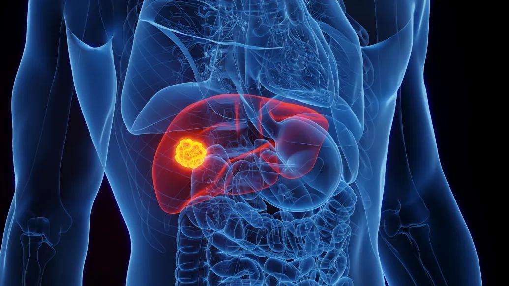
Some patients who have undergone abdominal imaging may have been alarmed to see a mention of a “benign-appearing cyst” of the liver in their results.
But these simple liver cysts are surprisingly common and mostly harmless.
An unexpected mention of a cyst in a patient portal, however, can be surprising.
Here, Robert J. Fontana, M.D., a Michigan Medicine hepatologist and a Professor of Internal Medicine, answers some questions you may have if you’ve received such a result after a CT scan, MRI or ultrasound.
What is a benign-appearing liver cyst?
Liver cysts are a common benign finding of the liver that are frequently detected on a liver ultrasound or CT scan of the abdomen done in patients with abdominal pain, vomiting or other gastrointestinal symptoms.
Simple cysts are small round or oval-shaped fluid-filled structures—usually less than 2 cm (1 inch) in diameter—that develop in the liver parenchyma and do not communicate with the biliary system.
The fluid arises from the lining cells of the cysts, and the cysts have a smooth edge without any solid components or calcification.
They are generally benign with a very low likelihood of enlargement or causing symptoms from rupture or malignant transformation.
How common are simple liver cysts?
Multiple studies show that up to 5% of adults have one or more small simple liver cysts.
In general, liver cysts are more common in women than men, with women being 50% more likely to have a cyst and their incidence increases with age.
What percentage deemed “benign appearing” turn out to be not benign?
The vast majority of simple liver cysts do not grow or enlarge during follow up. However, some patients have simple liver cysts in bad locations that abut against vascular structures or the bile ducts, which can cause problems via local compression.
In addition, complex cysts, which have irregular or thickened borders or a solid component, can infrequently grow or cause symptoms and may require biopsy or removal.
What causes simple cysts in the liver?
Liver cysts are believed to be congenital in nature in most individuals and are not detected until the liver is imaged in adulthood.
In contrast, complex cysts in the liver may develop from infections—like a hydatid cyst or prior bacterial infection—or tumors that arise from or spread to the liver.
Do simple liver cysts typically require treatment?
It's recommended that patients with one or more simple liver cysts that are less than 2 cm in maximal diameter do not require any treatment or follow up due to their low likelihood of symptoms or enlargement.
In contrast, the rare patients with genetically mediated polycystic liver and kidney disease or Carolis disease typically have dozens of large cysts in the liver that can communicate with the biliary tree and should be followed by a GI or liver specialist.
Patients with genetic polycystic liver disease should also avoid estrogen supplements.
Rarely, larger cysts that are at the surface of the liver can rupture, hemorrhage or become infected and may require radiological or surgical drainage.
Sign up for Health Lab newsletters today. Get medical tips from top experts and learn about new scientific discoveries every week.
Sign up for the Health Lab Podcast. Add us wherever you listen to your favorite shows.

Health Lab
Explore a variety of health care news & stories by visiting the Health Lab home page for more articles.
Media Contact

Public Relations
Department of Communication at Michigan Medicine
In This Story

Robert John Fontana, MD
Professor
Stay Informed
Want top health & research news weekly? Sign up for Health Lab’s newsletters today!
Featured News & Stories
IBS treatment response predicted by gut microbiome in new study
Low FODMAP diet improves leaky gut in study

FDA approves gastrointestinal device

How a phone call led Michigan Medicine to become a leader in treating severe ulcerative colitis

New guideline for Helicobacter pylori includes change to primary treatment recommendation