Revolutionizing head injury care
How a simple blood test is changing the game in emergency medicine
5:00 AM
Author |
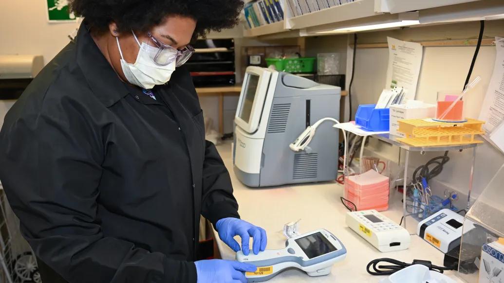
One Monday afternoon Francine Bomar was picking up her granddaughter from school when a sudden fall changed her day, and put a groundbreaking new Michigan Medicine emergency department innovation to the test.
“I was walking too close to the edge of the sidewalk,” Bomar said. “I went off the side and did a major face plant.”
At the ER, she feared long wait times and a CT scan.
Instead, she received rapid, compassionate care and something unexpected.
“I was checked in and was escorted to a unit that could take care of me immediately,” she said. “I couldn't stop thanking all the folks that took care of me. They were so kind and efficient in getting me out within the hour."
What stuck with her wasn’t just the care; it was the new blood test that helped her avoid a CT scan and get released quicker.
Novel traumatic brain injury testing
The new traumatic brain injury blood test, introduced at Michigan Medicine in December 2024, measures two brain-specific proteins —GFAP and UCH-L1.
Developed by Abbott Laboratories, the test is the result of years of research and validation by national experts in traumatic brain injury research, including Fred Korley, associate chair of research and professor of emergency medicine.
Korley has been studying TBI for nearly two decades.
“For a long time, I have been looking for a way to optimize how we care for head injuries,” said Korley.
“Nearly everybody with a head injury gets a CT, but it can be hard to determine who needs one or needs further care.”
Each year, about 5 million CT scans are performed in the United States for head injuries.
Waiting for one can take hours.
This new test, however, delivers results in 15 minutes and does so safely.
“It’s accurate and very sensitive to severe injuries,” said Katharine Seagly, M.D., clinical associate professor of physical medicine and rehabilitation, and program director for the TBI Rehabilitation Program.
Between the end of December 2024 and May 2025, Michigan Medicine avoided 104 brain CTs, Korley said.
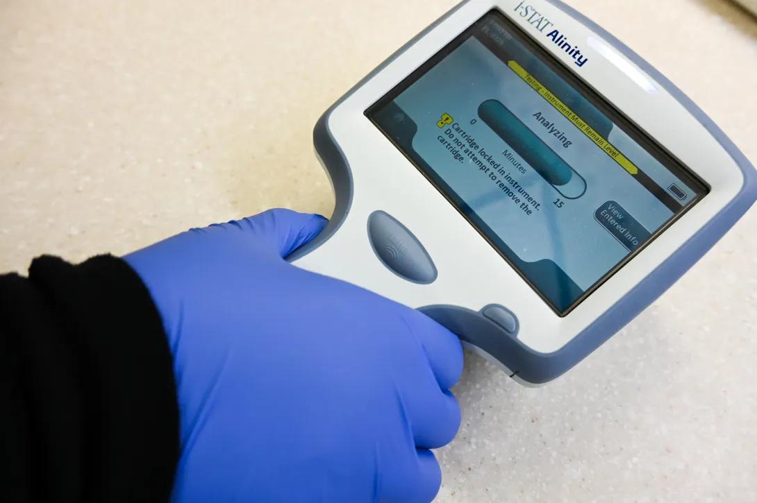
According to Nathan Haas, the associate medical director of the emergency critical care center and clinical assistant professor of emergency medicine, the test has both clinical and operational advantages.
“It’s exciting to think about the potential benefits for patients by avoiding ionizing radiation,” Haas said.
“It allows for a yes-no, algorithmic-type decision. And it helps with operational efficiencies in the ED."
That enthusiasm is echoed by others who’ve seen the impact firsthand.
“It’s a game changer for emergency medicine,” said Ben Bassin, Michigan Medicine associate medical director of survival flight and medical director of emergency critical care center.
“The ER is often in reactive mode, filling the gaps when others are closed, but this lets us be proactive. Unlike much research. This actually makes it to the bedside, and it works.”
Though the FDA approved it in 2018, Michigan Medicine is among the first in the country to use the test clinically.
“It’s exciting to be early adopters pushing the envelope,” Haas said.
Before launching it clinically, Korley and his team spent two years validating it through an internal study of 2,000 patients.
They compared blood test results with CT scans to fine-tune its application.
Early development of the device used for testing was supported by funding from the Massey Grand Challenge through the Weil Institute, which helped lay the groundwork for its clinical use.
This is one of the most important things I’ve done in my career so far ... It’s about balancing quality care with access to care. If current practice causes long waits, it needs to change. This is a safe way to offer the same level of care, improve access and reserve CTs for those who really need them.”
-Katharine Seagly, M.D.
Bomar agrees.
She is hopeful about the broader use of the test.
“How great it would be, cost-wise, to have this test available,” Bomar said.
She also reflected on how innovation can ease pressure on emergency departments.
“I hope it continues so that people with more serious injuries can be seen more quickly,” Bomar said.
Korley and his team are already looking ahead.
“Ultimately, we want to make prehospital decisions for patients and use this test to develop new, personalized treatment strategies for persons with traumatic brain injuries,” said Korley.
A grant co-led by Korley and Mark Burns, professor of chemical engineering, is helping them develop a handheld version of the test — about the size of a credit card — that could take 10 minutes or less, and be used in ambulances, nursing homes, on the battlefield, and elsewhere without CT access.
They're also working to integrate it into national care guidelines.
The grant, from the Department of Defense, will allow them to explore the test’s full potential.
“This test only reports on two biomarkers, but it could use 100s for a variety of things,” Korley said.
They plan to study 120 biomarkers to monitor secondary brain injury biomarkers, helping clinicians understand if an injury is worsening or improving.
“Our goal is for this to become part of the standard of care,” said Korley.
“Not just here at Michigan, but everywhere.”
Health Lab
Explore thousands of health news & research stories by visiting the Health Lab homepage for more.
Media Contact
Public Relations
Department of Communication at Michigan Medicine
In This Story

Frederick Korley, MD, PhD
Professor
Stay Informed
Want top health & research news weekly? Sign up for Health Lab’s newsletters today!
Featured News & Stories
What’s changing in Medicaid and what can you do?
Michigan Alzheimer's Disease Research Center receives $22M to advance efforts statewide

Rabies is on the rise: Experts offer advice
Engineered nanoparticles sensitize gliomas to radiotherapy

E-Bikes are everywhere, but most parents say they don’t know enough about them