Path forward for glioblastoma treatment
Experts discuss the latest brain cancer research
5:00 AM
Written by Rebecca Dzombak. This article was originally published on April 26, 2024 and updated on June 30, 2026.
While brain cancers are not always lethal, the most common form, glioblastoma, is aggressive and incurable, with a median survival of one and a half years.
From identifying deadly tumors sooner, understanding the biology of how and why they grow, and uncovering promising new treatments, researchers at University of Michigan Health Rogel Cancer Center are carving a path that creates hope for patients.
Two-pronged attack for high grade gliomas
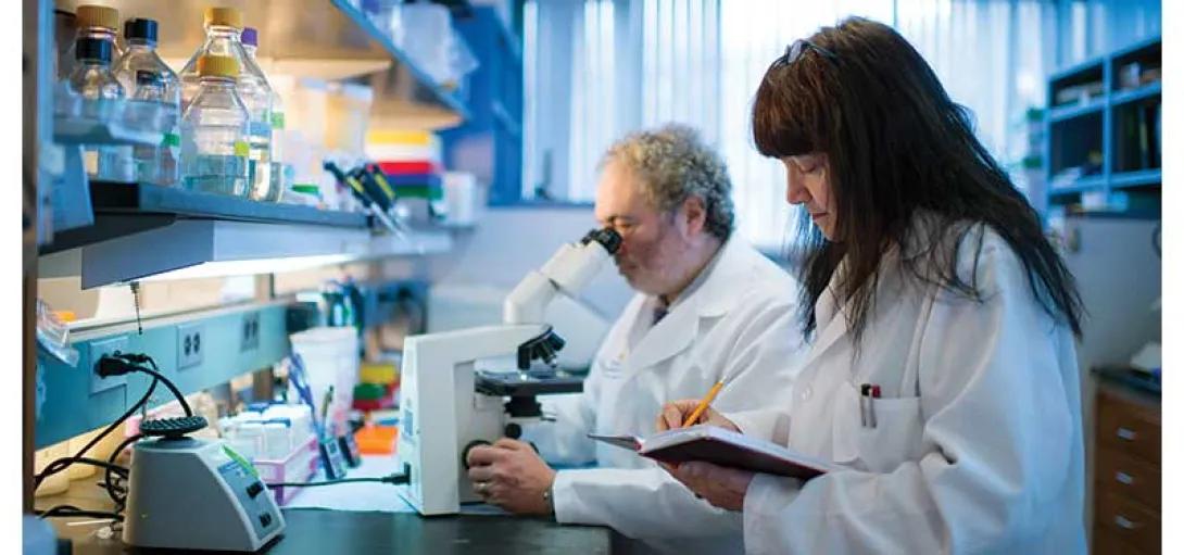
Maria Castro, Ph.D., and Pedro Lowenstein, M.D., Ph.D., are leaders in tackling aggressive brain tumors.
They specialize in gene therapy-mediated immunotherapy for high grade gliomas and collaborate with researchers across U-M to translate their therapies for adult and pediatric patients.
Their work, more than two decades in the making, is about to pay off.
“Gliomas are very aggressive, and it’s important to make progress, but that progress can be terribly slow,” said Castro, R.C. Schneider Professor of Neurosurgery, Professor of Cell and Developmental Biology and Co-Director of the Biosciences Initiative in Brain Cancer Technologies.
“There’s been a lot of progress in identifying gliomas, imaging them and running diagnostics, but this has had little impact on therapeutic outcomes.”
When Castro first began working with Lowenstein on brain tumors in the 1990s, gene therapy was a new, emerging field, and their team was at the frontier’s edge, focused on treating high grade gliomas.
“No one was doing gene therapy,” Castro said.
“It was a very new technology.”
Single-vector approaches were effective for killing rapidly dividing cancer cells, but sparking an immune response in the brain was challenging.
Lowenstein, Richard C. Schneider Collegiate Professor of Neurosurgery, Professor of Cell and Developmental Biology, and Professor of Biomedical Engineering discovered that the brain’s immune system lacks dendritic cells, which are crucial for initiating immune responses.
That lack could explain the difficulty in eliciting effective anti-glioma immune responses.
To trigger a natural immune response in the tumor cavity after resection, Castro and Lowenstein wanted to use a new two vector approach: “One gene kills the tumor cells, the other activates the immune system,” Castro said. “It’s a two pronged attack.”
Castro, Lowenstein, and their team built two adenoviral vectors to be injected into the brain cavity post-resection.
The early results were surprisingly promising, Castro and Lowenstein recall.
“When you use each therapy independently, neither one works so well,” Castro says. “But when you combine them, the efficacy just rises through the roof.”
The therapy also worked particularly well for model animals with gliomas that carried the IDH1 mutation, which Castro describes as “a death sentence delayed in time.”
Mutant-IDH1 tumors always come back and are resistant to radiation treatments.
But patients with mIDH1 tumors tend to have longer survival times—in part, as Castro recently published in Science Advances, because the mIDH1 tumor microenvironment is epigenetically modified such that the immunosuppressive cells have lost their immune suppressive capacity.
Castro and Lowenstein’s dual-vector approach takes advantage of that mutation.
In early studies, they found 98% of animals with mIDH1 glioblastomas were cured, a finding “never seen before in any tumor type,” Castro said.
“It worked beautifully.”
Castro and Lowenstein moved to Michigan Medicine in 2011 and began the study of the dual vector treatment, targeting high grade glioma patients with wild type IDH1 in 2013 with support from the Phase One Foundation.
The results, published in The Lancet Oncology, from 18 patients treated as part of a Phase I trial were stunning.
Patients in the study saw median survival times of about 21 months, a significant gain from the historic survival of 14.6 months.
The two year survival rate was nearly 40%; seven patients survived beyond two years, three survived beyond three years, and one was still alive 60 months after initial study enrollment.
“We believe this gene therapy approach may be even more powerful in patients with the IDH1 mutation,” Castro said.
“I predict it will be really effective in that patient population, so that’s one of the future trials we’re working on now.”
Part of the promise of the treatment is the potential for long term therapy. Castro and Lowenstein found that HSV1-TK persisted in patients’ brains for more than two years.
Currently, Valacyclovir is only authorized to be administered for a total period of four weeks, so their findings suggest that if the administration of Valacyclovir could be extended to months or years, patient survival times could be extended proportionally.
“This was totally unexpected,” Lowenstein said of HSV1-TK’s longevity.
“It gets me particularly excited, it’s very promising. It certainly justifies going into larger trials.”
Lowenstein and Castro are now developing a phase Ib/II trial for the dual-vector approach for wild type and mIDH1 glioma patients.
Overall, they hope their work will help the field and that the FDA will have broader acceptance of multi-pronged treatments like theirs.
“The way to push this forward is in combination,” Castro said.
Not only one diagnosis
Pediatric oncologist Andrea Franson, M.D., works with Castro and Lowenstein to bring their dual vector treatment to pediatric patients.
Franson’s focus has been on high-grade gliomas with the H3G34R mutation, which is present in a subset of patients with pediatric high-grade glioma patients.
This effort is supported by Ian’s Friends Foundation.
Typically, these patients live only 12 to 24 months from diagnosis.
“In so many of these tumor types, the need for better treatments and outcomes is so desperately high,” Franson said.
“We’re dealing with these really rare tumors, so thankfully the numbers of patients are low, but there are still a lot of tumors and a lot of need.”
Like mIDH1 tumors, pediatric H3G34R mutant tumors have incapacitated immunosuppressive cells, making them potentially susceptible to the dual-vector treatment.
Franson, Assistant Professor of Pediatrics, is working under the mentorship or Castro and Lowenstein to explore anti-tumor immune responses in H3G34-mutated versus non-H3G34-mutated tumors in mice, probing the system for points that could be exploited to trigger an immune attack using the dual-vector treatment.
Translating Castro and Lowenstein’s genetic immunotherapy approach for pediatric patients is critical, in part because so many tried and true treatments have fallen short.
“Chemotherapy, radiation therapy and surgery may work for the short term, but the tumors don’t stay away,” Franson said.
“People have tried many, many things over the last several decades, but for the toughest tumors, there’s really been no progress.”
Part of the delay in successfully treating H3G34R high-grade gliomas lies in researchers’ lack of knowledge about their fundamental biology.
Scientific understanding of brain tumors’ genetics (including histone mutations like H3G34R) didn’t come about until recent years.
“The more we dig into tumor genetics, genomics and proteomics, the more we realize there are all these subcategories of tumors,” Franson said.
"We may be giving patients the ‘same’ diagnosis, but really, it’s not.”
Franson hopes to hone treatment by improving tumor profiling at the diagnostic state.
While that reality may be distant, methylation profiling is a promising step down that pathway and may lead to different treatments choices made from the time of diagnosis.
Franson is a co-lead investigator for a national a phase I clinical trial that’s using genetic tumor profiling to optimize drug treatment.
“If we understand enough about how these tumors form and progress, we’ll get closer to finding what actually helps patients,” Franson said.
Pediatric oncologist Carl Koschmann, M.D., who serves as co-director of the Chad Carr Pediatric Brain Tumor Center, is developing a treatment for pediatric patients with diffuse midline gliomas with the H3K27M mutation.
The median survival time for patients with these high-grade gliomas is around 12 months, even with radiation therapy, “so there’s a lot of progress to be made,” Koschmann said.
“Currently, we can’t remove them. We can barely biopsy them.”
Koschmann’s dual life in the clinic and the lab, including starting his own lab in 2016 after training with Castro and Lowenstein, has enabled him to work on the cutting edge of both understanding and treating tumors with this mutation.
His most recent advancement builds on studies of ONC201, a dopamine receptor antagonist, that has been explored for use in diffuse midline glioma patients.
In an early study, ONC201 was found to be safe and cross the blood-brain barrier, but it was not effective at treating adult glioblastoma.
But several younger patients in that trial who had DMG that harbored a hallmark H3K27M mutation showed a potential response, opening the door to further study.
A phase I trial of ONC201 opened in 2017 to examine the H3K27M mutation and its tie to better survival times.
U-M was one of a few early trial sites.
During this time, Koschmann worked with Sriram Venneti, M.D., Ph.D., the co-director of the Chad Carr Pediatric Brain Tumor Center, to understand why the drug might be more effective in cells with the H3K27M mutation.
Their studies of spinal fluid from patients treated at U-M led to the discovery that ONC201 disrupts mitochondrial behavior and changes the tumor’s epigenetics, “essentially restoring tumor cells to be a lot more like normal brain cells after treatment,” Koschmann said.
Their study, published in Cancer Discovery, shows that in a cohort of 36 DMG patients treated after initial radiation, the median survival time was nearly 22 months.
This therapy is the first single therapy to improve outcomes for these patients, and a phase III trial is underway.
“Since 2017, we’ve treated more than 100 patients with this therapy here at U-M,” Koschmann said.
“Their overall survival is noticeably better than historical survival, so it’s exciting to see that there is a crack in the armor. I’m optimistic that in our generation of experimental treatments, we’ll have patients surviving five or 10 years, or beyond. That’s where the future is heading.”
Catch tumors early, treat them right
Because treatments for these brain cancers still offer relatively little to patients, catching the disease early—and maybe even predicting patients’ risks of developing them—is critical.
“Part of what makes these cancers so lethal and so hard to treat is a lack of early diagnostics,” Castro said.
"Think about skin cancer, where a lot of progress has been made. They recommend you do skin scrutiny once a year, and if you see something that looks suspicious, they immediately take it out. But with brain tumors, they’re silent killers. By the time the patient comes to the doctor, the tumor is already very advanced.”
Radiation oncologist Michelle Kim, M.D., is working to change that.
As professor of radiation oncology and co-chair of the Rogel Cancer Center neuro-oncology clinical research team, she’s developing more accurate, precise imaging to characterize and predict tumor growth early to inform individualized, targeted treatment.
Collaborating with Yue Cao, Professor and head of Functional Imaging in the Department of Radiation Oncology, Kim has worked to understand the imaging phenotypes characterizing the biologic features of glioblastomas and metastases.
Most recently, Kim focuses on using perfusion and diffusion-weighted multiparametric MRI to identify hyperfused and hypercellular regions in newly diagnosed, therapy-resistant glioblastomas.
The approach identifies regions that remain after resection and are missed by standard imaging technologies.
Targeting these regions of treatment-resistance using dose-escalated radiation therapy, a cohort of adult patients had favorable survival outcomes in a phase II trial led by Kim and her collaborators.
“The initial outcomes from this study appear to be promising,” Kim said, and the work is being developed into a national phase II/III randomized trial through the National Clinical Trials Network.
To support this effort, Kim received the inaugural NIH/NCI R50 Research Specialist (Clinician Scientist) Award in early 2023.
She's developing this study through the Alliance for Clinical Trials and NRG Oncology, which could be the first fully collaborative cooperative study between two major oncology groups in the United States.
“We are very excited about this potential opportunity to pioneer a new direction in the use and implementation of integral imaging biomarkers to improve outcomes in patients with malignant brain tumors,” Kim said.
Brain cancer research future
The need and the momentum for rapid, radical change in the field is clear to all these researchers, whether it’s detecting tumors sooner, throwing new combinations of treatments at the disease or building a national registry of brain cancers.
Despite the tough uphill battle, Rogel researchers remain dedicated and hopeful.
“At U-M and in the field of neuro-oncology, we recognize the importance of multidisciplinary collaboration,” Kim said.
“There’s not just one person who’s going to find the silver bullet. Collective creativity and thinking from different angles are critical for improving patient outcomes. And that’s the approach and the legacy we have here at U-M.”
“We need to work together, combine our efforts to attack this monster from several angles,” Castro added.
“Once we’re able to combine multiple approaches immunotherapy, radiation, chemotherapy, targeted gene therapies we’ll be able to make a dent in this disease. This is what we believe quite strongly.”
"We’re going to be here until we find a cure"
From 2014 to 2015, Koschmann treated Chad Carr, a 4-year-old boy diagnosed with DIPG.
When Chad passed away, the relationship that had formed between the Carr family and Michigan Medicine led to Chad’s mother, Tammi Carr, forming the ChadTough Foundation, which supports pediatric patients and their families and funds national research on DIPG and other aggressive, high-grade gliomas.
“When Chad was diagnosed, Michigan hadn’t focused much on DIPG,” Carr said.
"We wanted to do something to improve that, because that’s Michigan, that’s family.”
In under a decade, the ChadTough Foundation and its donors have helped Michigan become an international leader in pediatric brain cancer treatment and research, including the foundation of the Chad Carr Pediatric Brain Tumor Center in 2016.
“In such a short period of time, U-M is now without a doubt one of the top centers in the world when it comes to DIPG,” Carr said.
“Michigan has said, ‘We need to be better. We need to do better.’ ”
In 2020, ChadTough joined with the Michael Mosier Defeat DIPG Foundation, created in 2015 after Michael passed away from DIPG.
Together, the ChadTough Defeat DIPG Foundation has partnered with more than 40 families around the country to spread awareness, supporting patients and their families and funding critical research.
“Ultimately, we’re not where we want to be, but progress is being made, and life expectancy is basically increasing. There are more trials now than ever,” Carr said.
“We want every family to feel like their child is honored and remembered and making an impact on the disease. We’re going to be here until we find a cure.”

Health Lab
Explore a variety of health care news & stories by visiting the Health Lab home page for more articles.
Media Contact
Public Relations
Department of Communication at Michigan Medicine
In This Story
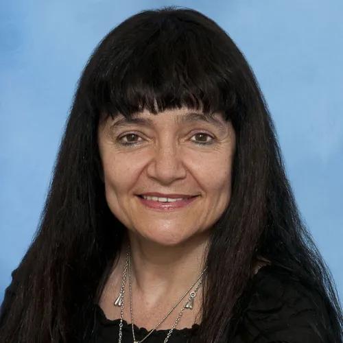
Maria Castro, PhD
Professor
Andrea (Flynn) Franson, MD, MS
Clinical Associate Professor
Pedro Lowenstein
Professor
Related
Turning the Tables on Glioblastoma
Stay Informed
Want top health & research news weekly? Sign up for Health Lab’s newsletters today!
Featured News & Stories

Researchers create new path to target hard-to-drug prostate cancer protein

How new care models, within a new building launch, are helping optimize patient care

Studying neurons using neurons
How AI is helping emergency physicians learn from their patients
Research may help better predict outcomes in kids with congenital cytomegalovirus