New prostate biopsy technique shows potential future of MRI
Transgluteal approach helps patients not served by fusion biopsies
1:30 PM
Author |
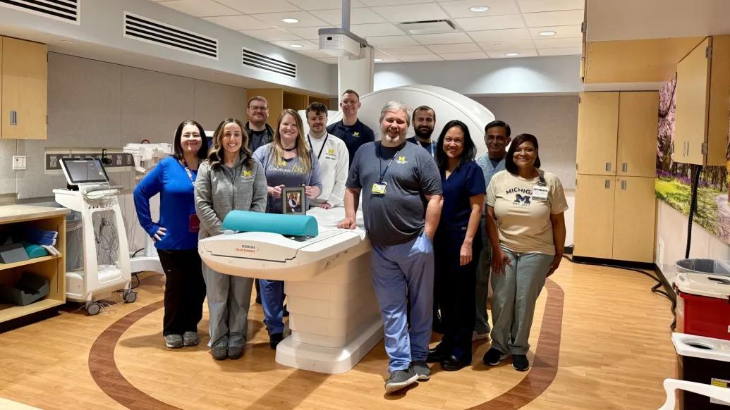
Michigan Medicine physicians and researchers are investigating new in-bore, MRI-guided prostate biopsy techniques, which can help patients not served by standard methods.
Specifically, University of Michigan Health radiologists have been the first physicians nationally to perform percutaneous transgluteal biopsies in a 0.55T MRI system.
Early published results have found the procedure to be effective.
Transgluteal biopsies use MRI imaging to guide the biopsy needle to the prostate through the gluteal muscle, as opposed to through the rectum.
This approach serves patients who lack a rectum — possibly because of past cancer treatment — or who otherwise cannot tolerate the rectal probe.
It also offers an alternate sterile route to the prostate, which does not require general anesthesia. (Percutaneous transperineal biopsies are also sterile but generally require anesthesia support.)
MRI also provides superior imaging ability to standard biopsy methods.
“The superior soft tissue contrast and real-time capabilities of MRI improve our ability to target suspicious lesions in the prostate and elsewhere, providing very high diagnostic capabilities and an opportunity to improve treatment outcomes with other percutaneous interventions, such as tumor ablation,” said Shane A. Wells, M.D., Associate Professor of Radiology and Urology.
The 0.55T MRI system is a scanner with a lower field strength and a larger opening for the patient to enter (80 centimeters in diameter as opposed to the typical 60-70 centimeters.)
Using a scanner of this size allows greater room for biopsy devices — which can be important in patients with larger habitus — and aids with patient comfort in the scanner.
“When the 0.55T came on, the first question was: Can you even use it for biopsy?” said Vikas Gulani, M.D., Ph.D., Chair of the Department of Radiology at U-M Health.
“The standard of care for diagnosing prostate cancer is typically 3T. Thus, most in-bore biopsy studies have also utilized the higher field strength. However, our data are very strong that, yes, you can use the 0.55T. You can see and target the lesions to be sampled, and you can do this accurately.”
For people whose initial tests show them at risk of prostate cancer, a prostate biopsy is an important diagnostic step.
Most prostate biopsies are fusion biopsies, in which an MRI image — already indicating a suspicious lesion — is combined with an ultrasound performed in a urologist’s office.
The urologist can then use the fused image of the two scans to accurately guide the biopsy needle transrectally.
While this approach is generally effective, it does not serve every patient, especially those lacking a rectum or whose rectum is diseased and thus cannot allow the instrumentation for biopsy.
In-bore biopsy is the most accurate way to perform a prostate biopsy. However, it is not practical to perform this for every patient, due to the large number of patients needing biopsy.
In addition to patients who cannot undergo the typical fusion biopsy, the transgluteal biopsy can be performed for problem solving in complex clinical scenarios, such as cases in which a fusion biopsy result and clinical or imaging suspicion do not match up.
The 0.55T magnet also reduces imaging artifacts caused by metal, such as prostheses or the biopsy needle itself.
“It’s possible for a patient to have a negative prostate biopsy despite blood markers indicating cancer, because a prosthetic was hiding a lesion,” said Elaine Caoili, M.D., M.S., the Saroja Adusumilli Collegiate Professor of Radiology at the University of Michigan Medical School.
“The 0.55T magnet sometimes allows us to see cancer that has been obscured on other images.”
Researchers believe that it will be possible to further develop the capabilities of MRI-guided interventions in the future, by taking advantage of some inherent abilities of MRI, which can combine the real time abilities of ultrasound with soft tissue contrast that is superior to CT scans.
As the speed of MRI increases, Michigan Medicine radiologists view these biopsies as a jump off point for more applications of the technology.
“Reduced artifact and a wider-bore design make the 0.55T system a promising platform for MR-guided interventions more broadly,” Yun Jiang, Ph.D., Assistant Professor of Radiology at the University of Michigan Medical School.
“The next step is to optimize sequences and workflow and determine how these advantages can be applied to other MR-guided procedures.”
Funding/disclosures: This research was partially supported by NIH grants R37CA263583 and R01CA208236, Siemens Healthineers, and Cook Medical.
Paper cited: “Clinical Feasibility of MRI-guided In-Bore Prostate Biopsies at 0.55T,” Abdom Radiol (NY). DOI: 10.1007/s00261-024-04783-x
Sign up for Health Lab newsletters today. Get medical tips from top experts and learn about new scientific discoveries every week.
Sign up for the Health Lab Podcast. Add us wherever you listen to your favorite shows.

Health Lab
Explore thousands of health news & research stories by visiting the Health Lab homepage for more.
Media Contact

Public Relations
Department of Communication at Michigan Medicine
In This Story

Elaine M Caoili, MD, MS
Clinical Professor
Stay Informed
Want top health & research news weekly? Sign up for Health Lab’s newsletters today!
Featured News & Stories
Immune system protein is a cancer killer on its own, new study shows
The AI is listening: Implications of machine generated clinical notes on health care

Comprehensive DNA sequencing reveals hidden drivers of cancer

Many older adults make Medicare decisions quickly and most don’t change plans, poll finds

‘Molecular glue’ stabilizes protein that inhibits development of non-small cell lung cancer and uterine cancer