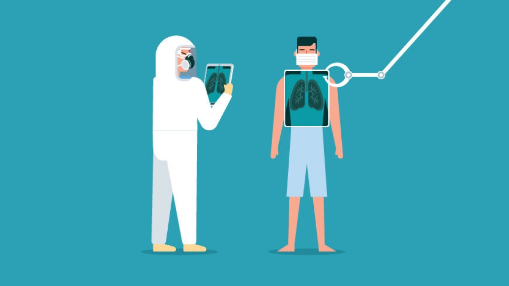
A Michigan Medicine radiologist teams up with a counterpart in China to examine the appearance of COVID-19 in CT scans.
11:00 AM
Author |
Editor's note: Information on the COVID-19 crisis is constantly changing. For the latest numbers and updates, keep checking the CDC's website. For the most up-to-date information from Michigan Medicine, visit the hospital's Coronavirus (COVID-19) webpage.
Interested in a COVID-19 clinical trial? Health research is critical to ending the COVID-19 pandemic. Our researchers are hard at work to find vaccines and other ways to potentially prevent and treat the disease and need your help. Sign up to be considered for a clinical trial at Michigan Medicine.
While COVID-19, previously known as the novel coronavirus, was first reported in China, it was recently declared a global health emergency by the World Health Organization. Because most cases have been in China, clinicians elsewhere may be unfamiliar with how the virus appears in the lungs.
And reports are now showing the importance of computed tomography (CT) in diagnosis and monitoring of the infection.
"As COVID-19 continues to evolve on a global scale, it is important for radiologists to be familiar with the imaging appearance of the virus in patients," says Prachi Agarwal, MBBS, M.D., a professor of radiology at Michigan Medicine. "Radiologic work here is extremely crucial when it comes to making diagnoses for patients."
This inspired Agarwal to team up with Weifang Kong, M.D., a radiologist at Sichuan Provincial People's Hospital in Chengdu, China, to examine the appearance of COVID-19 in three separate case studies involving patients with the condition.
Their research was recently published in Radiology: Cardiothoracic Imaging.
"Our work focused on the imaging appearance of COVID-19," says Agarwal. "The radiographic and CT appearance is not specific to the disease and can be seen with other infections, too."
Like Podcasts? Add the Michigan Medicine News Break to your Alexa-enabled device or subscribe for updates on iTunes, Google Play and Stitcher.
The pair discovered that while the imaging appearance of COVID-19 is not specific, the presence of bilateral nodular and peripheral ground glass opacities and consolidation should serve as an alert to radiologists that COVID-19 may actually be present in certain patients.
For example, individuals who have a defined travel history to areas where others are infected by COVID-19, or have been directly exposed to others with the virus, should be examined carefully.
As COVID-19 continues to evolve on a global scale, it is important for radiologists to be familiar with the imaging appearance of the virus in patients. Radiologic work is crucial when it comes to making diagnoses for patients.Prachi Agarwal, M.D.
Agarwal adds that in radiologic terms, 'ground glass' means that a hazy lung opacity shows up on imaging that is not dense enough to obscure any underlying pulmonary vessels or bronchial walls. While consolidation, on the other hand, refers to dense opacities obscuring vessels and bronchial walls.
Since ground glass opacities are common in COVID-19, Agarwal notes that chest CT scans are preferred over chest radiographs, which may have limited sensitivity in picking up early changes within the lungs.
"Chest CT scans can be helpful in suggesting the diagnosis for a patient and also, for monitoring patient responses," says Agarwal. "It's important to note that there is a spectrum of clinical manifestations and clinical course in COVID-19. And the clinical presentation of the virus can overlap with other respiratory illnesses."
MORE FROM THE LAB: Subscribe to our weekly newsletter
Some patients who tested positive for COVID-19 were either asymptomatic or had minimal symptoms. And while the reference standard for making the diagnosis is a real-time reverse transcription polymerase chain reaction (RT-PCR) test, false negative results can occur.
"It has been shown that an abnormal chest CT scan can predate a positive RT-PCR, highlighting the important role of CT in the management of these patients," says Agarwal.
To learn more about COVID-19, visit: What Can You Do to Protect Against Coronavirus?
Paper cited: "Chest Imaging Appearance of COVID-19 Infection," Radiology: Cardiothoracic Imaging. DOI: 10.1148/ryct.2020200028

Explore a variety of healthcare news & stories by visiting the Health Lab home page for more articles.
Department of Communication at Michigan Medicine
Want top health & research news weekly? Sign up for Health Lab’s newsletters today!