A multi-institutional study finds that COVID-19 can be found in post-mortem corneal tissue, highlighting the importance of the donor screening process.
11:40 AM
Author |
COVID-19 has been found in conjunctival swabs and tears of infected patients, according to a new study published in The Ocular Surface.
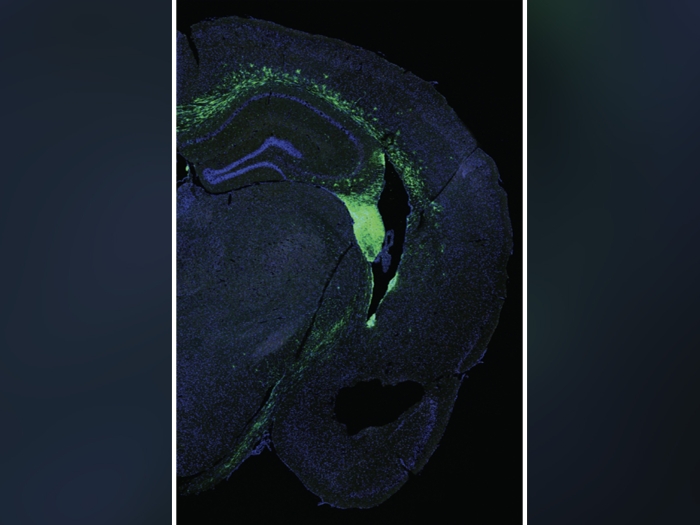
The discovery prompted a research team including Shahzad Mian, M.D., an ophthalmologist at Kellogg Eye Center, to analyze the prevalence of COVID-19 in human post-mortem ocular tissues. The results: the virus can infiltrate corneal tissue, the clear, outer layer of the eye, that could be used for transplantation in the U.S., raising concerns that the disease could be transmitted to a healthy recipient.
Of the 132 ocular tissues from 33 donors intended for surgery in Michigan, Illinois, Ohio and New Jersey, 13% were positive for COVID-19, which was determined by isolating the ribonucleic acid (RNA), a molecule similar to DNA, of the patients that were known to have the virus or showed symptoms without a positive nasopharyngeal swab.
LISTEN UP: Add the new Michigan Medicine News Break to your Alexa-enabled device, or subscribe to our daily updates on iTunes, Google Play and Stitcher.
Studies have shown that COVID-19 patients hold much of the virus in the upper respiratory tract, so there's a strong possibility the virus could contaminate the outer layers of the eye via respiratory droplets after coughing, sneezing or hand-to-eye contact, according to Mian.
"There's no evidence to suggest COVID-19 can be transmitted from a corneal transplant, but our data assures us that a screening process to determine who's positive for the virus and who isn't is important to make sure we do everything in case there is a potential risk of transmission," Mian adds.
These questions are important in keeping our patients healthy and safe.Shahzad Mian, M.D.
The findings also demonstrate the critical importance of post-mortem nasopharyngeal swab testing for detecting COVID-19 before transplantation. The study's donors were divided into three groups:
- Group 1
This group was positive for COVID-19 after receiving a nasopharyngeal swab at the time of corneal recovery.
- Group 2
This group was primarily made up of donors from early in the pandemic when testing wasn't widely available. The majority of these donors had a negative COVID-19 test.
- Group 3
This group didn't have signs or symptoms of COVID-19 and tested negative, but they also spent extended amounts of time with someone who tested positive.
This is significant because 15% of the corneal samples from Group 2 presented with COVID-19 RNA, despite having a negative nasopharyngeal swab test. In fact, this was even higher than the presence of coronavirus-infected corneal tissue from Group 1, which only had a positivity rate of 11% despite the donors having positive nasopharyngeal swab tests.
None of the tissues from Group 3's two donors had a presence of COVID-19 RNA.
Lowering the presence of COVID-19 in the cornea
Aside from establishing a screening process, Mian set out to discover if there was a way to lower the presence of COVID-19 in the donor tissue -- another strategy that could diminish transmission risk.
"An initial study goal was to test the effectiveness of povidone-iodine, a disinfectant, in inactivating COVID-19," Mian says.
This was done by recovering donors' right eyes without cleaning with povidone-iodine and following the Eye Bank Association of America recommended double povidone-iodine soak procedure on the left eyes.
The procedure involves soaking the cornea in 5% povidone-iodine for five minutes, then flushing with sterile saline fluid. This is repeated after a sample of corneal tissue is harvested. All of the eyes that underwent this disinfecting tested negative for COVID-19 RNA, compared to one of the right eye swabs that tested positive.
MORE FROM MICHIGAN: Sign up for our weekly newsletter
However, the team is unable to conclude that povidone-iodine is at all effective in reducing COVID-19 in corneal tissue because this procedure was only performed on 10 patients.
"A larger study is needed to confirm our findings, but we're excited about this research's potential implications. These questions are important in keeping our patients healthy and safe," Mian says.
It's unclear whether the presence of COVID-19 RNA is due to ocular surface infection or due to transport of the virus from the upper respiratory tract via the tear ducts. It's also unclear whether COVID-19 can replicate in corneal cells and what changes occur in these cells when infected.
"The takeaway I hope other physicians have when reading this study is that following elaborate donor screening procedures to mitigate the risk of pathogen transmission during transplant, as well as testing post-mortem donors for COVID-19 specifically, when there is no COVID-19 nasal swab testing, is critically important as professionals in this field."
The Eversight Center for Vision and Eye Banking Research, Wayne State University and Rush University contributed to this ongoing research. This study is supported by Eye Bank Association of America, National Eye Institute and National Institute of Allergy and Infectious Disease.
Paper Cited: "Prevalence of SARS-CoV-2 in human post-mortem ocular tissues," The Ocular Surface. DOI: 10.1016/j.jtos.2020.11.002

Explore a variety of healthcare news & stories by visiting the Health Lab home page for more articles.
Department of Communication at Michigan Medicine
Want top health & research news weekly? Sign up for Health Lab’s newsletters today!