An AI model that can read and diagnose a brain MRI in seconds
Researchers say the technology has potential applications beyond neurological diagnoses
5:00 AM
Author |
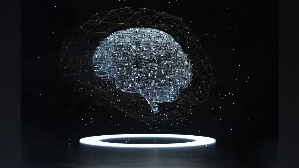
An AI-powered model developed at University of Michigan can read a brain MRI and diagnose a person in seconds, a study suggests.
The model detected neurological conditions with up to 97.5% accuracy and predicted how urgently a patient required treatment.
Researchers say the first-of-its-kind technology could transform neuroimaging at health systems across the United States.
The results are published in Nature Biomedical Engineering.
“As the global demand for MRI rises and places significant strain on our physicians and health systems, our AI model has potential to reduce burden by improving diagnosis and treatment with fast, accurate information,” said senior author Todd Hollon, M.D., a neurosurgeon at University of Michigan Health and assistant professor of neurosurgery at U-M Medical School.
Hollon calls the invention Prima. He and his research team tested the technology on more than 30,000 MRI studies over the course of a year.
Across more than 50 radiologic diagnoses from major neurological disorders, Prima outperformed other state-of-the-art AI models on diagnostic performance.
The model also succeeded in determining which cases should take higher priority.
Some neurological conditions, such as brain hemorrhages or strokes, require immediate medical attention. In such cases, Prima can automatically alert providers so rapid action can be taken, Hollon says.
Researchers designed the model to recommend which subspecialty provider should be alerted, such as a stroke neurologist or neurosurgeon, with feedback available immediately after a patient completes imaging.
“Accuracy is paramount when reading a brain MRI, but quick turnaround times are critical for timely diagnosis and improved outcomes,” said Yiwei Lyu, M.S., co-first author and Ph.D. student of Computer Science and Engineering at U-M.
“At key steps in the process, our results show how Prima can improve workflows and streamline clinical care without abandoning accuracy.”
What is Prima?
Prima is a vision language model, an AI system that can simultaneously process video, images and text in real time.
It’s not the first attempt to apply AI to MRI and other forms of neuroimaging, but the approach is unique.
Past models rely on manually curated subsets of MRI data to achieve specific tasks, such as detecting lesions or predicting dementia risk.
When designing Prima, Hollon’s team trained the system on every MRI — over 200,000 studies and 5.6 million sequences — taken since radiology digitization began University of Michigan Health decades ago.
“Our teams at University of Michigan have collaborated to develop a cutting-edge solution to this problem with tremendous, scalable potential.”
-Vikas Gulani, M.D., Ph.D.
Researchers also input patients’ clinical histories and the physicians’ reasons for ordering medical imaging study into the model.
“Prima works like a radiologist by integrating information regarding the patient’s medical history and imaging data to produce a comprehensive understanding of their health,” said co-first author Samir Harake, a data scientist in Hollon’s Machine Learning in Neurosurgery Lab.
“This enables better performance across a broad range of prediction tasks.”
Millions of MRI studies are performed globally each year, with a significant portion focused on neurological diseases.
This demand, researchers say, outpaces the availability of neuroradiology services and leads to significant challenges, including workforce shortages and diagnostic errors.
Depending on where you get a scan, it can take days, or even longer, to get a result.
“Whether you are receiving a scan at a larger health system that is facing increasing volume or a rural hospital with limited resources, innovative technologies are needed to improve access to radiology services,” said Vikas Gulani, M.D. Ph.D., co-author and chair of the Department of Radiology at U-M Health.
“Our teams at University of Michigan have collaborated to develop a cutting-edge solution to this problem with tremendous, scalable potential.”
The future of AI and imaging
While Prima performed well, the research is in its initial stage of evaluation.
The research team’s future work will explore integrating more detailed patient information and electronic medical record data for more accurate diagnosis. This strategy closely emulates how radiologists and physicians interpret MRIs and other radiology studies.
Health care providers, systems and policymakers are still determining how to appropriately integrate artificial intelligence into practice, yet most systems currently used are for narrow medical tasks.
What Hollon describes as “ChatGPT for medical imaging” has broader potential — and could one day be adapted for other imaging modalities, such as mammograms, chest X-rays and ultrasounds.
“Like the way AI tools can help draft an email or provide recommendations, Prima aims to be a co-pilot for interpreting medical imaging studies,” Hollon said.
“We believe that Prima exemplifies the transformative potential of integrating health systems and AI-driven models to improve health care through innovation.”
Additional authors: Asadur Chowdury, M.S., Soumyanil Banerjee, M.S., Rachel Gologorsky, Shixuan Liu, Anna-Katharina Meissner, M.D., Akshay Rao, Chenhui Zhao, Akhil Kondepudi, Cheng Jiang, Xinhai Hou, Rushikesh S. Joshi, M.D., Volker Neuschmelting, M.D., Ashok Srinivasan, M.D., Dawn Kleindorfer, M.D., Brian Athey, Ph.D., Aditya Pandey, M.D., and Honglak Lee, Ph.D., all of University of Michigan.
Funding/disclosures: This work was supported in part by the National Institute of Neurological Disorders and Stroke (K12NS080223) of the National Institutes of Health.
The content is solely the responsibility of the authors and does not necessarily represent the official views of the NIH.
This work was also supported by the Chan Zuckerberg Initiative (CZI), Frankel Institute for Heart and Brain Health, the Mark Trauner Brain Research Fund, the Zenkel Family Foundation, Ian’s Friends Foundation and the UM Precision Health Investigators Awards grant program.
Michigan Research Core(s): UM Advanced Research Computing
Paper cited: “Learning neuroimaging models from health system-scale data,” Nature Biomedical Engineering. DOI: 10.1038/s41551-025-01608-0
Sign up for Health Lab newsletters today. Get medical tips from top experts and learn about new scientific discoveries every week.
Sign up for the Health Lab Podcast. Add us wherever you listen to your favorite shows.
Health Lab
Explore thousands of health news & research stories by visiting the Health Lab homepage for more.
Media Contact
Public Relations
Department of Communication at Michigan Medicine
In This Story

Todd C Hollon, MD
Associate Professor

Vikas Gulani, MD, PhD
Professor
Related
Artificial intelligence predicts genetics of cancerous brain tumors in under 90 seconds
In 10 seconds, AI model detects cancerous brain tumor often missed during surgery
Stay Informed
Want top health & research news weekly? Sign up for Health Lab’s newsletters today!
Featured News & Stories
A pediatric epilepsy fundraiser that changes lives
Better prenatal care data could help prevent maternal complications, experts say
Pain, psychological factors and physical function closely linked in people with systemic sclerosis
Many hospitals still don’t do enough to prevent UTIs from catheters

Digital alerts support timely treatment decisions of critically ill cardiac patients