An interdepartmental team at the University of Michigan obtains a prestigious NIH grant that will support an advanced imaging strategy to improve methods for early cancer detection.
5:00 AM
Author |
Worldwide, the incidence of cancer is steadily growing. Research has shown time and time again that this rise is often connected to the prevalence of obesity, coupled with an increasingly aging population, and society's regular consumption of a high-fat diet.
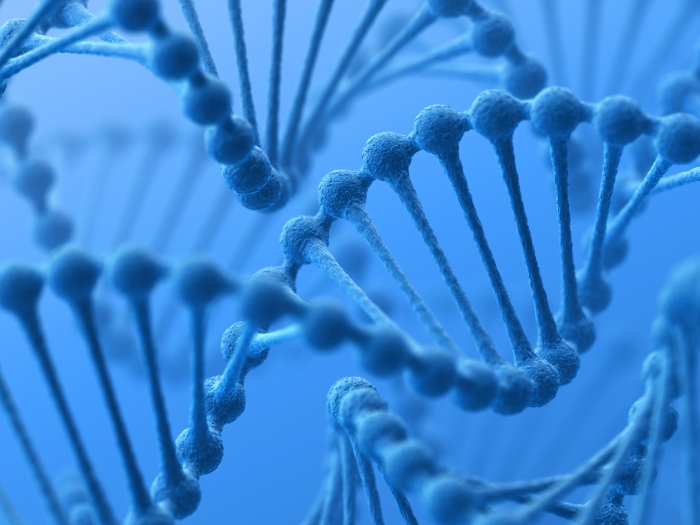
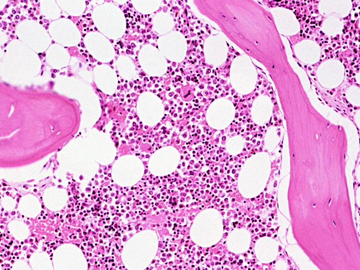
Over 80% of all human cancers originate from the epithelium of hollow organs and ducts. This thin layer of tissue is characterized by a simple, yet repetitive structure, that holds intense self-renewing capabilities and is an incredibly important site in the world of early cancer detection.
These notions intertwined to inspire Thomas Wang, M.D., Ph.D., H. Marvin Pollard Collegiate Professor of Endoscopy Research, Jason Spence, Ph.D., associate professor of internal medicine and cell and developmental biology and Kenn Oldham, Ph.D., associate professor of mechanical engineering, to apply for and obtain a $3 million grant from the National Institute of Biomedical Imaging and Bioengineering (NIBIB) branch of the National Institutes of Health (NIH).
"This grant will support a cutting-edge imaging strategy to advance improved methods for early cancer detection in the epithelium of human organs and ducts," says Wang. "We're very excited about this initiative, which will show that some of the most capable tiny devices ever developed can be applied to make a real impact on cancer diagnosis and treatment," says Oldham.
Endoscopy, then and now
Research has shown a "miss rate" for identifying and removing colon polyps of greater than 20%, even while the early detection and prevention of colorectal cancer is regularly performed via screening colonoscopies.
Even further, flat and often depressed non-polypoid dysplasia make up 25% of all polyps and are often harder to detect via colonoscopy, while possessing higher risks for malignancy. However, improvements in image contrast when viewing these pre-malignant lesions may ultimately result in better detection and mortality rates, as well as better patient outcomes, overall.
"Molecular imaging is a powerful tool for both detecting and guiding therapy for the early stages of cancer," says Wang. "It has the power to assess protein expression patterns on the surface of cells transforming into malignancy. This is really powerful stuff."
Together, we have pioneered an aggressive and dynamic leveraging mechanism to enable robust, large-angle scanning of light for extremely compact tools for use during endoscopy.Thomas Wang, M.D., Ph.D.
Often, these molecular changes occur prior to any detectable cell abnormalities. Yet, by detecting non-polypoid dysplasia through wide-field imaging techniques in real-time, there are better chances for localizing these pre-cancerous lesions to help physicians remove them.
"The proposed wide-field imaging methods will serve as a supplement to improve conventional endoscopy methods for the early detection of colorectal cancer," says Wang. "Other technologies include narrow-band imaging, chromoendoscopy and autofluoresence imaging, which all have their own unique limitations."
LISTEN UP: Add the new Michigan Medicine News Break to your Alexa-enabled device, or subscribe to our daily updates on iTunes, Google Play and Stitcher.
This is why narrow-band imaging, for example, which allows diseased tissue to appear darker in color than surrounding healthy mucosa, can't aid in the detection of dysplasia alone, and still requires white light endoscopy for guiding tissue biopsy. Chromoendoscopy utilizes non-specific exogenous dyes, such as indigo carmine, to enhance micro-anatomical changes in the mucosa.
However, like that imaging, chromoendoscopy relies on white light endoscopy to guide tissue biopsy and may miss flat lesions that don't present sufficient contrast from topographical changes of the epithelium alone. Auto-fluorescence imaging is highly sensitive to endogenous fluorescence from other molecules, like collagen. Debris and mucosal folds can cause shadows, inhibiting the accuracy of this approach.
"We know that in order for our approach to be effective, it must improve upon the current imaging capabilities," says Wang. "And through a collaborative approach, our initiative aims to accelerate the development of promising technologies to address early cancer detection and other problems in medicine through advancing integrative, quantitative and engineering methods."
MORE FROM THE LAB: Subscribe to our weekly newsletter
A trifold plan
The team, led by Wang, Oldham and Spence, will consist of engineers, chemists, biologists and physicians. The program will involve three key components:
-
Oldham will use high-work density materials to develop tiny scanners using microsystems technology at U-M's Lurie Nanofabrication Facility to generate the images in real-time.
-
Wang will develop a flexible, yet miniature, fiber-based imaging instrument that will be used as an accessory to standard medical endoscopes when performing in vivo imaging of the epithelium in hollow organs and ducts.
-
Spence will implant human organoids in the colon of mice to verify instrument performance. These lesions are pre-malignant and have a flat appearance that make detection with endoscopes challenging. The organoids express cancer-associated genes at levels representative of what are seen in human patients.
"Together, we have pioneered an aggressive and dynamic leveraging mechanism to enable robust, large-angle scanning of light for extremely compact tools for use during endoscopy," says Wang. "This is very exciting."

Explore a variety of healthcare news & stories by visiting the Health Lab home page for more articles.
Department of Communication at Michigan Medicine
Want top health & research news weekly? Sign up for Health Lab’s newsletters today!