4 times you might need an ultrasound and didn’t know
Ultrasound’s little-known uses include drug injections and MRI alternatives
5:00 AM
Author |

Do you need an ultrasound?
Before you answer, consider that you—and even your primary care physician—might not be aware of everything ultrasound imaging can achieve these days.
Improvements in technology have increased the resolution of images.
And with a top team to record and interpret your images—like the University of Michigan’s Division of Musculoskeletal Radiology—ultrasound can help in ways you might not expect.
Below are four uses of the technology that U-M radiologists want to put on your radar.
1.Using an ultrasound during an injection
If you need an injection at the site of an injury, pain, or arthritis, the diagnosing physician might be eager to perform it right away. But the naked eye can’t monitor the vital structures the needle will pass through.
Even if an office has an ultrasound machine, the person administering the injection will not always be trained in image-guidance.
“I do think that most injections should always be performed under ultrasound-guidance,” said Steven Soliman, D.O., associate professor, program director of the Musculoskeletal Radiology Fellowship, and the director of Musculoskeletal Ultrasound at the University of Michigan.
“There's no radiation involved. It's low cost. And there are many small structures like nerves and vessels that could easily be injured without ultrasound-guidance. We monitor these important structures in real-time when we are closely watching the needle as it enters the exact area we are targeting.”
Injections performed without image-guidance might also not be as effective as one administered under the supervision of a radiologist.
The Musculoskeletal Radiology team at Michigan has encountered patients whose previous treatments did not provide much relief as the needle may have failed to find the affected area when performed without image-guidance.
2. Using an ultrasound instead of an x-ray
When a joint hurts, a doctor might order an x-ray.
Injuries to the soft tissues around those joint bones, however, will show up better on an ultrasound.
“I am not sure if the patients and their clinicians are always aware that the musculoskeletal ultrasound can be ordered and performed while waiting to see the specialist,” said Heidi Ehrich, RDMS, RMSKS, the lead musculoskeletal sonographer for Michigan Medicine.
Similarly, an ultrasound can discover many of the issues that might otherwise show up on an MRI at a fraction of the patient’s cost or time.
Ultrasounds can also provide answers before a specialist is available to diagnose and order an MRI.
3. Using an ultrasound to determine what’s clicking or popping
Not all physical problems are apparent when the patient is still. But other imaging methods take static images of patients lying down.
Ultrasound, which takes dynamic images, is uniquely suited to capturing a tricky injury associated with a specific movement.
If someone has a shoulder that pops when they swing their tennis racket—or a knee that hurts when they walk—a trained musculoskeletal radiologist can observe the problem area dynamically as their patient demonstrate movement.
“There are unique cases in which Orthopedics will actually send patients back to us for a dynamic ultrasound and after they’ve had an MRI,” Soliman said.
“They want us to evaluate real-time on ultrasound how an elbow ligament or ankle tendon functions dynamically prior to surgery.”
4. Using an ultrasound when an MRI seems unfeasible
The ability of ultrasound to substitute for MRI in some cases is good news for patients who are claustrophobic—or otherwise physically incapable of lying in a confined space for an extended period.
Sometimes a patient’s insurance might not be willing to cover for the MRI right away and will require an ultrasound first.
Other times, an MRI might be contradicted by a patient’s implant devices—or the quality of the image is degraded by devices or surgical hardware.
In those cases, ultrasound can be a great alternative.
“In such situations—as in all situations—the quality of the ultrasound matters,” Ehrich said.
“Experienced, board-certified musculoskeletal sonographers and radiologists, as we have here at Michigan Medicine, can help provide the accurate diagnosis or injection treatment that is really so important.”
Sign up for Health Lab newsletters today. Get medical tips from top experts and learn about new scientific discoveries every week.
Sign up for the Health Lab Podcast. Add us wherever you listen to your favorite shows.

Health Lab
Explore thousands of health news & research stories by visiting the Health Lab homepage for more.
Media Contact

Public Relations
Department of Communication at Michigan Medicine
In This Story

Steven B Soliman, DO, RMSK, FAIUM, FAOCR
Clinical Associate Professor
Stay Informed
Want top health & research news weekly? Sign up for Health Lab’s newsletters today!
Featured News & Stories
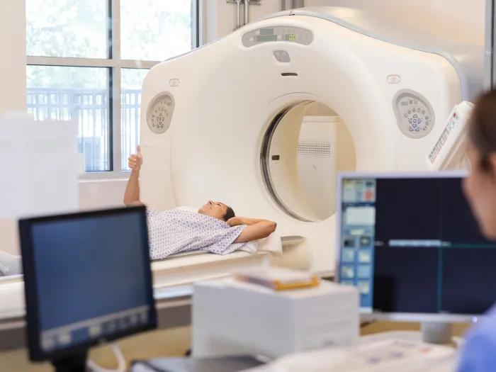
Hospital partnership improves follow up scans, decreases long term risk after aortic repair

An ultrasound-activated hydrogel for steady, sustained drug delivery

A way to measure brain blood flow in pre-term babies at the bedside

An AI model that can read and diagnose a brain MRI in seconds

AI model helps diagnose often undetected heart disease from simple EKG