Using biolasers to improve cancer diagnostic tools
Lighting up cancer cells with biolasers
5:00 AM
Lighting up cancer cells with biolasers. The technique overcomes the limitations of current cancer diagnostic tools. Read the full article on the Health Lab website.
Transcript
Host:
Welcome to Health Lab, your destination for news and stories about the future of health care. Today: Lighting up cancer cells with biolasers; using biolasers to improve cancer diagnostic tools.
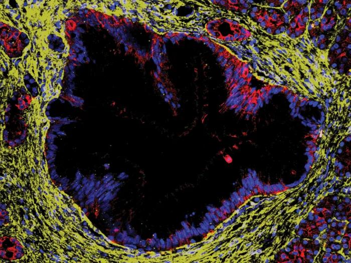
Researchers from the University of Michigan have developed a way of detecting circulating tumor cells in the bloodstream of pancreatic cancer and lung cancer patients.
The study was published in Biosensors and Bioelectronics.
As tumors develop, they shed cells into the bloodstream.
Although these circulating tumor cells are vastly outnumbered by millions of other blood cells, detecting them early can potentially improve treatment outcomes.
For instance, pancreatic cancer has a poor prognosis because by the time it has been detected, it is too late to treat.
Similarly, the detection rates of lung cancer, especially if it reoccurs after treatment, is also poor.
Current detection techniques for circulating tumor cells involve labeling specific proteins on the surface of tumor cells with fluorescent dyes.
These stained cells can then be easily detected in the blood samples.
However, there are some disadvantages.
Some of the cells may not have those proteins on their surface and can therefore be missed.
The techniques also miss valuable information about what’s happening inside the cancer cells.
“These existing techniques usually involve methods that end up killing the cancer cells, thus preventing us from utilizing these cells for further investigation,” said Sunitha Nagrath, professor of chemical engineering.
“We realized that we needed an alternative way to identify circulating tumor cells while they are alive.”
To do so, the researchers turned to biolasers.
Although the method still involves staining the cancer cells with dyes, it doesn’t kill them and instead of depending on proteins that are on the surface of the cells, the researchers can stain something that is integral to all cells—their nucleus.
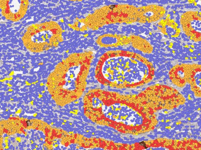
Using blood samples from patients with pancreatic cancer, the researchers first passed all the cells through a circular maze, called Labyrinth, that pre-separated out the circulating tumor cells, which are slightly larger than other white blood cells.
“It’s like driving around a curve in a bicycle versus a truck—the forces you experience are very different. As a result, the larger tumor cells get focused into different location compared to smaller white blood cells” Nagrath said.
They then sandwiched the tumor cells between two mirrors and shone an excitation laser at them one cell at a time.
When the excitation is strong enough, the cells have laser emissions and are referred to as cell lasers.
“The laser emission from a cell laser is much stronger than what we get from traditional fluorescent techniques,” said Xudong (Sherman) Fan, professor of biomedical engineering.
“The laser emission images are also different; in fluorescence emission the cells look like glowing spheres. However, with a laser you can see different shapes that provide information on how the DNA is organized inside cancer cells.”
These differences, however, are subtle.
The researchers therefore turned to machine learning for help.
Using the Deep Cell-Laser Classifier model, they were able to accurately pick out pancreatic cancer cells 99% of the time.
The model was so effective that even though it was trained using pancreatic cancer cells, it was able to identify lung cancer cells without requiring any additional training.
“Although there are a few research groups who are working with biolasers, we are the first to use it for clinical studies on cancers and circulating tumor cells,” Fan said.
Moving forward, the group is interested in building a device that can isolate cancer cells after they have been detected.
“With our system, if you want to collect circulating tumor cells, you have to remove the top mirror, which can cause the cell to move and then you lose track of it,” Fan said.
“We want to develop a system where cells move along one-by-one through the laser excitation spot and then go through a cell sorting device that helps us sort and collect cells for subsequent analysis.”
The team also plans to use the light patterns generated by the cells to better understand which tumors are more aggressive or treatment resistant.
“All these circulating cells can be very different from each other,” Nagrath said. “Identifying how aggressive cells change during treatment cycles would be helpful.” They go on, “This work would not have been possible without such an interdisciplinary team. The Judith Tam ALK Lung Cancer Research Initiative was a critical partner in enabling analyses of patient material. The resources we have are the stuff of dreams, and we are fortunate that we can use them to implement our innovative engineering ideas for clinical use.”
For more on this story and for others like it, visit michiganmedicine.org/health-lab where you can subscribe to our Health Lab newsletters to receive the latest in health research and information to your inbox each week. Health Lab is a part of the Michigan Medicine Podcast Network, and is produced by the Michigan Medicine Department of Communication. You can subscribe to Health Lab wherever you listen to podcasts.

Health Lab Podcast
Listen to more Health Lab podcasts - a part of the Michigan Medicine Podcast Network.
Featured News & Stories
Path forward for glioblastoma treatment
Study explains how colorectal cancer cells maintain high iron levels

The Race to Uncover the Hidden Causes of Idiopathic Pulmonary Fibrosis

Stopping Ewing sarcoma relapses where they start

Giving with gratitude: planned gifts support nursing, research, and patient care