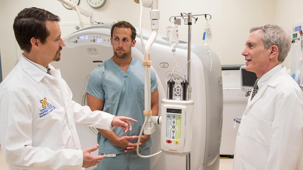
Matthew S. Davenport, M.D. (Residency 2010), assistant professor of radiology and of urology, is one of the leaders of a new effort to improve cancer diagnosis at U-M and throughout Michigan.
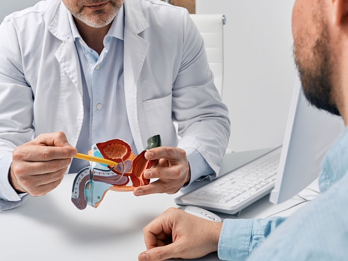
Historically, prostate-specific antigen, or PSA, and digital rectal examination, or DRE, have been the primary methods of detecting prostate cancer at a curable stage, but these methods are not ideal. Patients with clinically significant cancers can be missed, and patients with clinically insignificant cancers can be over-treated. Davenport and his collaborators in nuclear medicine, pathology and urology have been researching innovations in MRI, PET and ultrasound-MR fusion techniques to refine prostate cancer diagnosis for several years. And it has recently become available clinically at the U-M Comprehensive Cancer Center.
"In the past, if a patient's PSA was elevated, or they had an abnormal DRE, they would get a template biopsy directed to regions of the prostate rather than a defined target," Davenport says. "MRI allows more precise detection, targeted biopsies, and the ability to better predict whether the cancer is low-risk or aggressive."
U-M was the first in the region to offer this technique and the service is expanding rapidly. Prostate cancer care providers across the state are motivated to incorporate this technology into their own programs, thanks to its promising outcomes. Davenport and his urology colleagues are partnering with radiologists and urologists at other sites, and working with the Michigan Urological Surgery Improvement Collaborative to educate providers about advanced imaging, correct reporting of prostate MRI examinations and imaging-related data collection.
Davenport is also internationally recognized for his research on the safety and efficacy of radiographic contrast media that improve the visibility of pathology on CT and MRI examinations. He has co-authored national policy statements regarding contrast media, is director of body MRI at U-M and chair of the Michigan Radiology Quality Collaborative.